
Some canine fractures are so severe they require the expertise of a specialist in bone surgery. We have a specialist in bone surgery that will come to our hospital and perform the repair in dogs, cats, and other species like this rabbit. This has several advantages, not the least of which it costs less than if we refer the repair to a surgical specialist at a referral hospital.
This is a severe fracture, and a dog with this problem is very painful and can go into shock. This is a medical emergency requiring immediate veterinary care. The Long Beach Animal Hospital, staffed with emergency vets, is available until the evenings 7 days per week to help if your pet is having any problems, especially shock, pain, breathing hard, or bleeding.
Think of us as your Long Beach Animal Emergency Center to help when you need us for everything from minor problems to major a major emergency. We serve all of Los Angeles and Orange county with our Animal Emergency Center Long Beach, and are easily accessible to most everyone in southern California via Pacific Coast Hwy or the 405 freeway.
If you have an emergency that can be taken care of by us at the Animal Emergency Hospital Long Beach always call us first (562-434-9966) before coming. This way our veterinarians can advise you on what to do at home and so that our staff and doctor can prepare for your arrival. To learn more please read our Emergency Services page.
These pictures show the repair of Dakota, a Labrador who fractured his tibia (shinbone) by playing.
What is Fracture of the Tibia?
A tibia fracture is a broken bone. It can range from a slight crack in the bone to a complete break where the bone is in one or more segments.
Type of Fracture
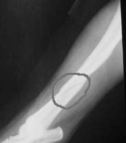
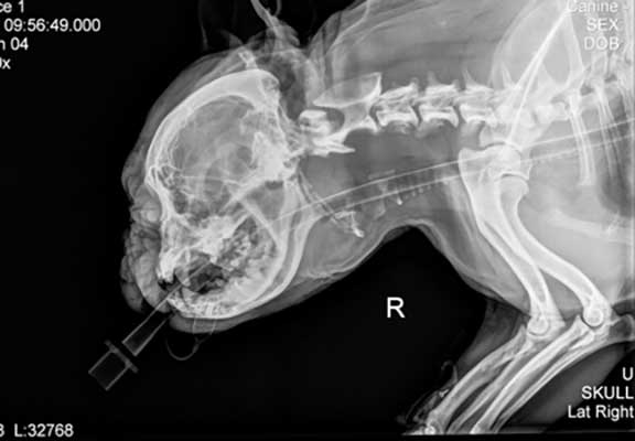
This fracture is called a spiral fracture due to the winding nature of the crack. The fracture is much more severe than is apparent on this x-ray. What is not apparent on the x-ray are the numerous bone fragments that were found surgically.
This is a severe fracture, so we have our orthopedic specialist Dr. Paul Cechner perform the surgery. He has decades of experience and the latest high tech equipment to do it right.
Symptoms of Fracture of the Tibia in Dogs
Symptoms can be as mild as slight whimpering or licking at the fractured leg, to limping and complete inability to use the leg.
Causes of Canine Tibia Fracture
In almost every case trauma is the cause. This is usually from falling from a height or being hit by an object like a ball, bat, or moving vehicle.
Diagnosis
Fractures of the tibia are routinely diagnosed with a radiograph. Even though a fracture can be suspected based on history and exam findings, a radiograph (x-ray) is needed for confirmation.
Graphic surgery photos on this page
Surgery Preparation
Our patient is given a thorough pre-anesthetic exam within 24 hours of his surgery.
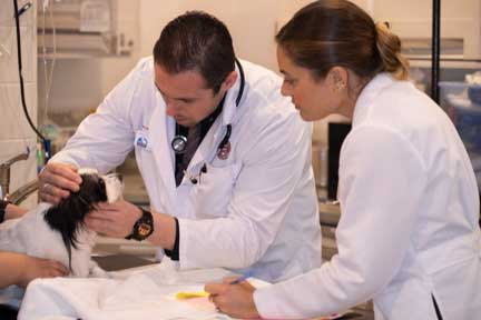
One of our student externs learning how to do a pre-anesthetic physical exam from Dr. Wood
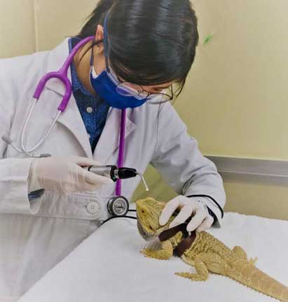
Even reptiles get get a pre-anesthetic exam
After the exam there are routine pre-anesthetic tests we perform. The three most important ones are a blood panel, radiographs, and a pre-anesthetic EKG (ECG).
Pre-surgical Preparation
Blood Panel
A dog that has trauma hard enough to break a leg might have some internal injuries that are not apparent externally when we perform a physical exam. One test to help us look for any problems is a blood panel.
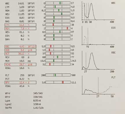
This is one page or our multiple page in-house blood panel. This pet is anemic, so we will be on the alert for internal bleeding
Radiology
This is especially important on a trauma case like this. Our patients do not talk to us, and they have high pain thresholds compared to us humanoids, so they don’t always show symptoms to let us know something else is a problem besides the fracture. We do not want any surprises on the day of surgery.
When we take a pre-anesthetic radiograph we are specifically looking for fractured ribs, pneumothorax, and a diaphragmatic hernia. This is important to minimize the risk of anesthesia, and to make sure all problems are identified and corrected.

This is a normal chest radiograph on a dog
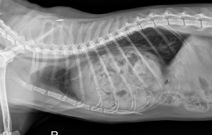
This is what a diaphragmatic hernia looks like. Can you see it compared to the radiograph above?
Learn much more about radiology from our Learn to Read an X-Ray page.
Pre-Anesthetic ECG
Animals with broken bones from being hit by a car (HBC) can have trauma to the heart that is not apparent when we listen with the stethoscope. For this reason we perform an electrocardiogram just prior to surgery.
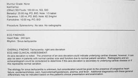
This one has a potential problem that needs to be addressed
In orthopedic surgery like this one we are inserting wires, bone plates, and screws. They will stay in long-term unless there is a problem with infection or rejection. Because of this sterility and aseptic technique is crucial, and it starts with our surgeon scrubbing his hands.
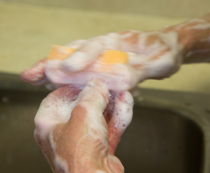
He thoroughly scrubs from his fingernails to his elbows

As soon as our surgeon is gloved and gowned he goes go right into the surgery suite to get the instruments ready. We do not want our patient waiting for our surgeon, we want the surgeon waiting for the patient.
Anesthesia
Once we have analyzed our pre-anesthetic exam and tests we will proceed to anesthetize our patient. They are given pre-anesthetic tranquilizers and pain medication to calm them down. Next they are given intravenous fluids.
Our Anesthesia Page has much more information on this.
Injectable Anesthesia
Injectable anesthetics are used for many purposes. One of their primary uses is to sedate pets before giving the actual anesthesia (called pre-anesthetic). In sedating ahead of time we dramatically minimize anxiety, cause a smoother recovery, and minimize how much anesthetic we need to administer during the actual procedure. In addition, some injectable anesthetics minimize vomiting, a common problem when waking up from anesthesia.

Injectable anesthesia is given intravenously, and rapidly induces relaxation so that we can put in a breathing tube (also called an endotracheal tube or ET tube)
Endotracheal Tube
Oxygen is delivered to the lungs by the ET tube in its windpipe. It is the preferred method to administer oxygen because it is very efficient, will prevent any vomitus from entering the trachea (vomiting rarely happens because of fasting and pre-anesthetic sedation), and allows us to gently inflate the lungs during surgery so that they work at maximum efficiency.
Besides oxygen, the anesthetic gas (Isoflurane) is also administered through the endotracheal tube. Medications can even be administered via this special tube.

The endotracheal (ET) tube is placed directly into the windpipe
This x-ray shows the breathing tube as it passes over the tongue and down the trachea (windpipe)
We can easily breathe for your pet and inflate your pet’s lungs by gently squeezing the bag connected to the tube, monitoring the amount of pressure we are exerting with a gauge on the anesthetic machine. Each size and species of pet requires a different sized endotracheal tube.
The tube is not removed from your pet until it is literally waking up. This ensures that the swallowing reflex is present, and your pet is now safely able to breathe on its own.
Gas Anesthesia
The mainstay for general anesthesia is gas anesthesia because it is very safe and highly controllable. We use a safe and effective gas anesthesia called Isoflurane. It is so safe it can be used in creatures as small as tiny birds. As soon as this patient is intubated it is put on gas anesthesia.
An instrument called a precision vaporizer is used to deliver the Isoflurane anesthetic gas within the oxygen. It is a very precise instrument allowing us to make fine adjustments in anesthetic level. Without this vaporizer we would not have the wide safety margin that we currently enjoy.

We can precisely and easily change the level of anesthesia during the procedure as needs change
For most surgeries we administer the anesthetic at a setting of 1-3 %. This small percent of anesthetic, added to the 100% oxygen the pet is breathing, is all that is needed to achieve complete surgical anesthesia. Before the surgical procedure is finished the anesthetic is lowered before it is turned off completely. As the surgeon is finishing the procedure your pet is in the beginning stages of waking up. This decreases anesthetic time, another way we minimize anesthetic risk.
Oxygen
All pets put under gas anesthesia are given 100% oxygen from the moment they are anesthetized until they wake up, dramatically increasing the safety of the procedure.

We have a special machine in surgery that generates 100% oxygen
As a backup, oxygen is stored in large tanks under high pressure. The oxygen in these tanks is delivered to the anesthetic machine via special piping throughout the hospital. This allows us to have anesthetic machines in several hospital locations.
Our patient is anesthetized and the leg is shaved outside of our surgery room. We do all of this outside of the surgery room to keep it as clean and sterile as possible.
Monitoring Anesthesia
Our surgeon is one of the best monitors, because he is literally visualizing the blood in the circulatory system. Any change in the blood is readily noticed because pets that are breathing 100% oxygen should have bright red blood.
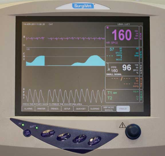
We use a sophisticated monitor to keep track of important physiologic parameters during surgery
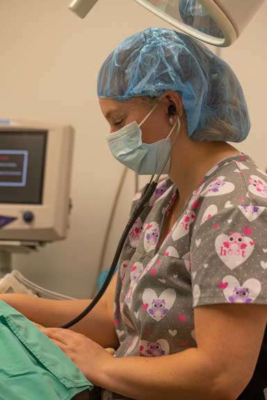
Even with all of that high tech monitoring equipment we still stay hands-on during the whole procedure

We keep detailed records of fluid rate rates, anesthetic and oxygen levels, and physiologic parameters, during the surgery
We have a very detailed page that shows you the wide variety of animals we anesthetize. Click here to learn much more about anesthesia.
Graphic surgery photos to follow
Surgery
The following area contains graphic pictures of an actual surgical procedure performed at the hospital. The surgery takes two hours, the following photos are just a summary of what it is like to fix a bad fracture on a large and active dog. We need to put them back together properly or the fracture will not heal.
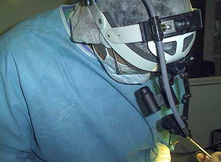
Our surgeon needs to utilize specialized equipment if he is to put this bone back together so that Dakota can return to normal function. In this picture he is using magnifying glasses and special lighting. In addition, he has orthopedic instruments and equipment without which he would never be able to repair such a severe fracture.
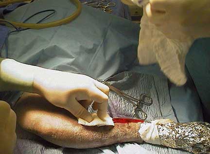
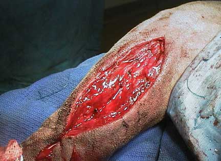
Bone infections can be serious so significant time is spent in sterile preparation. When Dakota has been anesthetized, and adequately prepared, an incision is made on the inside of his leg. This area has minimal muscle over it and gives good exposure to the fracture site.
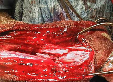
After carefully dissecting through muscle tissue over the bone the fracture is exposed
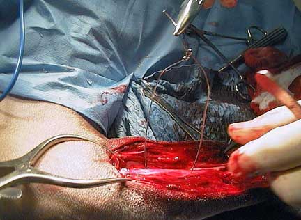
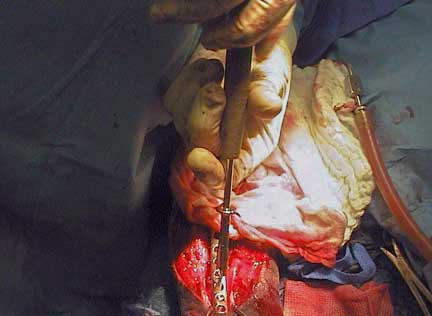
Once the bone fragments are isolated our surgeon uses special wires called cerclage wires to begin the process of holding the fracture segments in place. It is a tedious process that takes up a significant amount of the surgery.
The wire is tightened down with a special instrument that gives just the right amount of tension. Too little tension and the wire is useless, too much and the bone fractures even more.
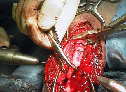
At this point 2 cerclage wires have been applied to the fractures at the top, with new ones being applied to the fractures at the bottom
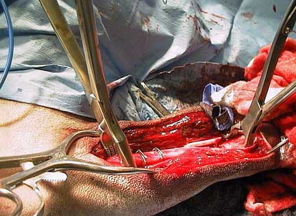
Eventually 6 cerclage wires are applied to align the bone fragments. Even though these wires are strong the bone will not stay in place and heal with just these wires. A bone plate is needed for most of the stability.
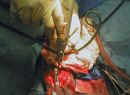
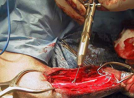
After the bone plate is measured and bent to the specific shape of this tibia, holes are drilled into the bone with a special air powered drill. They have to be drilled to the proper depth and angle or the bone will fracture more or the plate will fail.
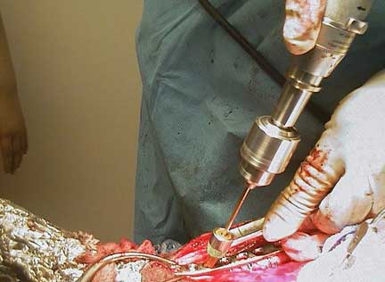
A special tap is used for proper placement
Drilling the holes is the first step in the application of the plate. The depth of the holes is measured, and specific screws are used. Some screws compress the plate to the bone, others hold the plate in place.

Two hours from the start of the surgery the plate has finally been applied. We will not remove it unless there is a post operative complication.
The muscle is sutured to preserve its function and to cover the plate. These sutures will slowly dissolve over several months.
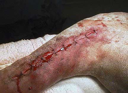
The skin sutures will stay in for 2 weeks. At this point in the surgery Dakota is given an antibiotic injection along with a pain injection. After one nights rest in the hospital he will go home. He will need to be confined for one month for healing to progress.
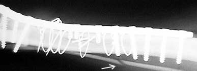
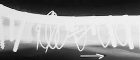
Before Dakota is fully awake from anesthesia an x-ray is taken to assess the surgery. The bend to the plate can be seen, along with the cerclage wires and the different lengths of the various screws. The fractured fibula (arrow) will heal by itself.
Once our surgeon is satisfied that everything is in order Dakota is given a pain injection and awakened from anesthesia. He will spend the night with us so that he can rest and so we can monitor his recovery. He will need to rest at home for several months before the healing is complete. We will not take the plate out unless complications arise.
How long does it take a tibia fracture to heal in dogs?
A slight crack in the bone can heal in 2-4 weeks, a complete fracture, especially that needs surgery, can take 3 months or longer to completely heal.