
This page confirms the importance of routine yearly exams, and close owner observation, on our pets. Large breed dogs can be stoic, and have significant problems brewing without showing any outward signs. When serious symptoms like weight loss and lack of appetite finally show up, the disease process is usually well entrenched, and there is little we can do. This can happen with an enlarged spleen in dogs. We want to see these pets for an exam at the Long Beach Animal Hospital before it gets to that stage.
Pets are masters at hiding illness, so it can be difficult to determine if they are ill. It might be a good idea to read our short page on symptoms of disease to know what to watch for. Don’t miss the case summary at the end of this page to learn more.
We also have a nice page on teaching you how to do an in-home exam. If you bring your pet to us one of our staff will go over this with you, and do a hands-on demonstration of how to palpate the peripheral lymph nodes. This early warning system is a great way to catch problems early in the course of disease.
A rupturing spleen is a medical emergency requiring immediate veterinary care. These dogs can collapse and go into life-threatening shock. The Long Beach Animal Hospital, staffed with emergency vets, is available until the evenings 7 days per week to help if your pet is having any problems, especially shock, seizures, pain, difficulty breathing, or bleeding.
Think of us as your Long Beach Animal Emergency Center to help when you need us for everything from minor problems to major a major emergency. We serve all of Los Angeles and Orange county with our Animal Emergency Center Long Beach, and are easily accessible to most everyone in southern California via Pacific Coast Hwy or the 405 freeway.
If you have an emergency that can be taken care of by us at the Animal Emergency Hospital Long Beach always call us first (562-434-9966) before coming. This way our veterinarians can advise you on what to do at home and so that our staff and doctor can prepare for your arrival. To learn more please read our Emergency Services page.
History
A beautiful male labrador retriever named Colt came in for a routine Wellness Exam when he was 10 years old. During this exam Colt’s owner told Dr. Palazzolo that Colt vomited on occasion over the last month, but that otherwise he was doing fine. He is a calm dog, and just lays around a lot.
Exam
Colt’s body temperature was normal, and his weight was the same as a previous visit. A pet’s weight is a good idea of how well it is doing, and if it is maintaining its weight that is usually a good sign. In Colt’s problem, this is not the case.
During the physical Dr. P noted a very tense abdomen, so tense that he could not even palpate internal organs. After this was noted, the owner did say that Colt’s abdomen seemed to be distended. The rest of Colt’s thorough physical examination was normal.
The distended abdomen is a significant finding, and warranted a radiograph, in addition to the blood panel which is part of the Wellness Exam.
Diagnostic Tests
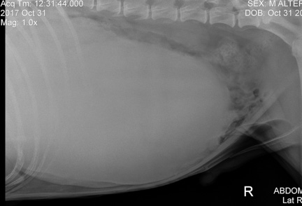
You don’t need to be a radiologist to know something is wrong with this abdomen, as evidenced by the large and round whitish structure
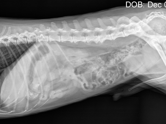
Here is a normal abdomen for comparison. In this normal radiograph you can see individual organs, unlike the abnormal one above, that looks like it has a soccer ball inside of it.
This is a serious finding, and this mass could have many causes, and involve almost any internal organ. Due to its size, shape, location, and the relative lack of symptoms, it was probably the spleen. It could be a benign tumor of the spleen called a hemangioma, a malignant tumor of the spleen called a hemangiosarcoma, or a hematoma of the spleen. A hematoma is a blood filled cavity that is benign.
Its size said it might be a hematoma, which has the best prognosis of the three. To learn much more about spleen tumors please visit our hemangiosarcoma page.
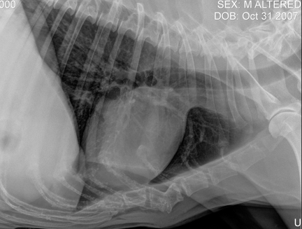
Before we proceed any further we take a radiograph of the chest to make sure there has not been any spread of tumor to the lungs. The white object in the center is Colt’s heart, the dark areas around the heart are the lungs. This is a normal chest radiograph.
You can learn much more about how to read a radiograph if you are interested.
Our radiologist, Dr. Ann Reed, performed an ultrasound of the abdomen. This ultrasound lets us know if the mass is actively bleeding, and if there has been any spread of the mass to the right atrium of the heart. If that is the case, the mass is probably a hemangiosarcoma that has already spread. This is not a good prognosis.

Colt’s ultrasound report showing it is probably benign, and consistent with a hemangioma or hematoma
Pre-surgical Preparation
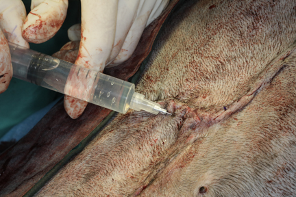
In Colt’s case there was no abdominal bleeding, and the heart was OK. Colt’s blood panel came back normal, so we did a cross match of his blood, and readied a unit of blood for a transfusion in case we needed it during the surgery scheduled for the next day. Before we do a blood transfusion we do several tests to minimize a transfusion reaction.
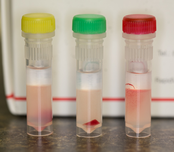
Some of the reagents we use to test the donor and recipient blood for a reaction
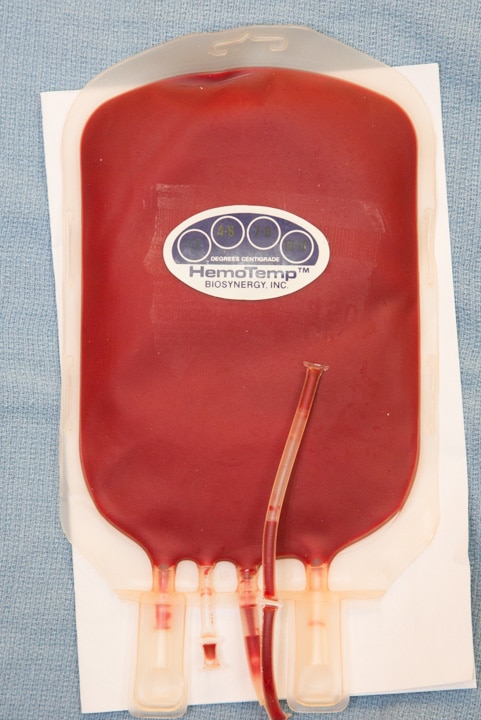
A fresh unit of whole blood ready if needed
We were much better prepared for surgery the next morning with all of this information. During the night prior to surgery Colt was given intravenous fluids to stabilize him and make him a better anesthetic risk, and a pain patch was put on. Having the pain medication on board before surgery increases its effectiveness. Colt is now good to go for surgery the next morning.
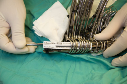
We have a short page that talks all about how we do surgery at the Long Beach Animal Hospital. You might want to check it out before you see Colt’s surgery.
Graphic and bloody photos of surgery to follow.
Pre-anesthetic preparation
Colt did great overnight. We took away his popcorn after the Lassie movie ended (he likes classic movies), and he rested comfortably during the night, monitored by our night crew. He slept well, and dreamed of being a famous movie star like Lassie.

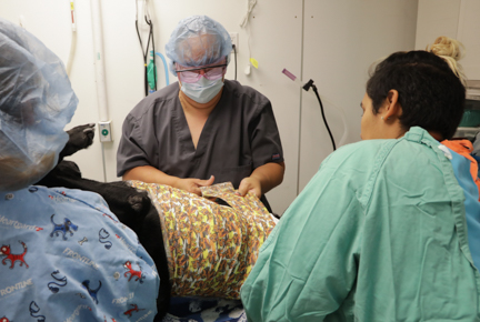
Here he is with Jennifer the next morning, keeping an eye on our surgical team as they prepare for his surgery

Colt gets some extra petting for being a good boy, by his surgeon, Dr. Kennedy
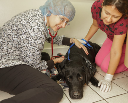
Dr. Kennedy doing her pre-anesthetic exam
Even though Colt has been thoroughly checked, we always perform a physical exam just prior to inducing anesthesia. An important organ to assess is the heart. If we suspect something is wrong with the heart when we listen to it we will do a pre-anesthetic EKG (electrocardiogram).
If you like heart stuff we have a very detailed page on heart disease in animals. It’s not for the “weak of heart” (pun intended), because it goes into all sorts of anatomy and physiology. If you get through it give yourself a pat on the back, and think of applying to veterinary school!
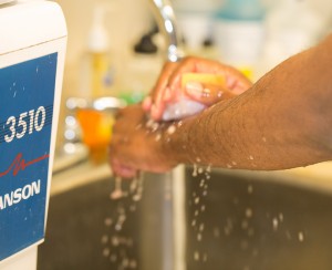
Once our surgeon has reviewed all lab data, and has performed the pre-anesthetic physical exam, she starts her aseptic scub while our patient is being anesthetized
After scrubbing and gowning our surgeon opens the sterile instrument pack to make sure everything is in order and all instruments are sterile
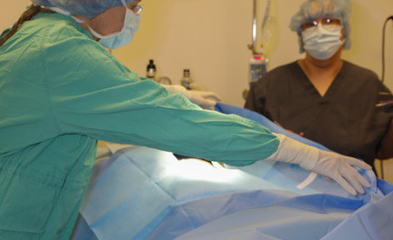
While all of this is transpiring with our surgeon our patient is brought into the surgery suite and a final prep is performed. We want our surgeon waiting for her patient, not the other way around. All of this is to minimize anesthetic time.
Anesthesia
When everything is to our satisfaction we will administer a sedative. This will calm her down and make the administration of the actual anesthetic, along with post operative recovery, much smoother. Many pets with Fentanyl patches do not need any additional sedative.
We keep a close tab on important physiologic parameters for all of our surgeries. Monitors like this give us an early warning of an impending problem. This machine monitors:
Temperature
Heart Rate
Heart rhythm
Oxygen saturation
Carbon dioxide level
Respiratory rate
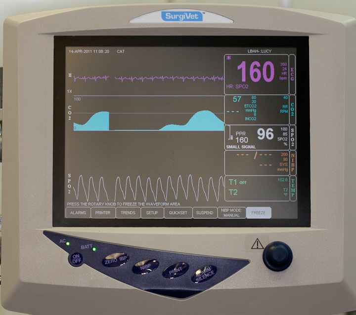
This is the screen we constantly monitor during surgery
The loud background sound in this video is our oxygen generator

Colt’s anesthesia is initially given by injection. Once he is relaxed we put in a breathing tube and start prepping his abdomen for surgery.
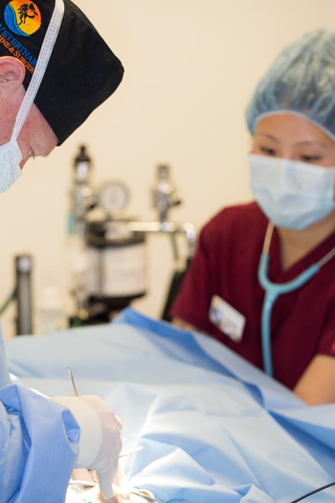
Even though we use these hi-tech machines to monitor important physiologic parameters during surgery, we also use the hands-on approach to make sure we do not miss anything. In this picture our anesthetist is listening to the heart with a stethoscope.
She also checks something called Capillary Refill Time to make sure the heart is pumping enough oxygenated blood to the organs. When she presses on the gums to make them blanch white for a second, she measures how long it takes them to go back to their original pink color. It should be less than two seconds.
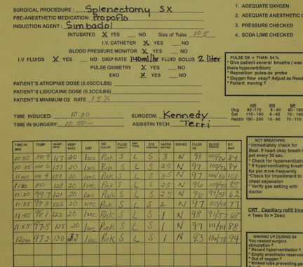
During the procedure Colt’s vital information is closely monitored and recorded
We have a detailed page on anesthesia to learn much more on how we anesthetize a wide variety of different species (like the duck below) at at the Long Beach Animal Hospital.
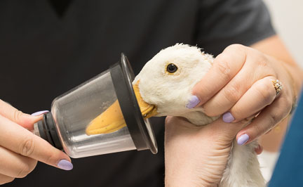
Modern anesthetics allows us to safely anesthetize high risk animals like this duck with a fractured wing. You can see her surgery in our Wildlife Care page.
Surgery
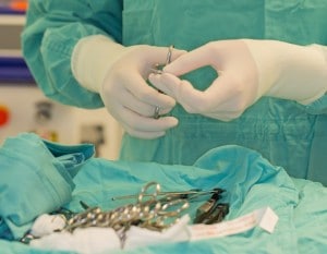
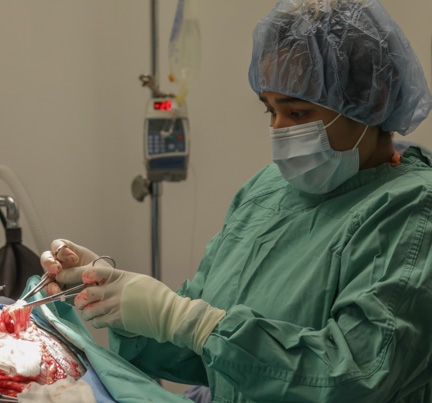
While Colt is being readied for surgery Dr. K is getting her instruments ready. She will need many clamps to close off the blood supply to the spleen before it can be removed, which required more than one surgical pack.
When Colt is prepped, and under the proper plane of anesthesia, Dr. Kennedy does her draping. This draping is an important part of the surgery, and is doing carefully.
It will take more than one pair of hands to get this spleen out, so our surgical assistant, Jessica, stands by waiting for instruction. Terri our anesthetist keeps a close tab on everything.

The initial incision is only though the skin. Blood vessels just under the skin are clamped before we go any further.
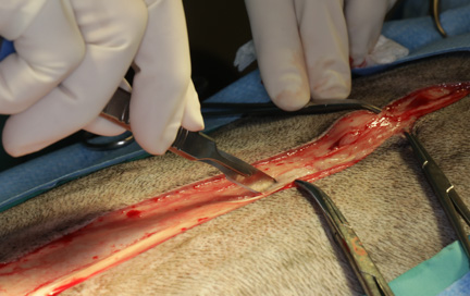
You can see the two clamps that are controlling bleeding in the subcutaneous (SQ) tissue just under the skin. Colt’s spleen is humungous, so it will take a long incision to get a big enough opening to remove it

Even though the incision was made as long as possible, the opening was still not big enough to remove the spleen. Dr. K had to make a side incision in the abdomen to facilitate removal.
The spleen has an extensive supply of blood vessels that are near the stomach. A large part of this surgery is identifying and ligating these vessels, and doing this without damaging the blood supply to the stomach. These blood vessels are intertwined in tissue that is covering the spleen as the body attempts to wall off this large mass. This is the most meticulous part of the surgery, and Dr. K’s experience and skill at doing this before pays off.

Identify a blood vessel to the spleen that is covered in fat

These blood vessels are large and need to be handled carefully
It takes multiple clamps and special suture material to perform this important part of the surgery
Jessica is kept busy holding clamps while Dr. K identifies and continues to ligate the many blood vessels to the spleen

As more of these vessels are ligated Dr. K carefully attempts to get the spleen out of the abdomen. No luck the first time.

An adhesion from the intestines to the spleen was identified and removed. Time to try to get the spleen out again.

We need to be very careful at this point that we do not rupture the spleen by forcing it out
With gentle coaxing and manipulating, along with Jessica’s help holding the abdomen open, Dr. K almost has it out, but not just yet

It was time for Dr. P to stop taking photos and assist in surgery. He has removed many spleens in his decades of surgery, and with the help of Dr. K and Jessica, was able to extricate the spleen. Now Dr. K could finish ligating blood vessels (and hopefully Jessica does not need to scratch an itch at her nose)!

Here she is doing a great job

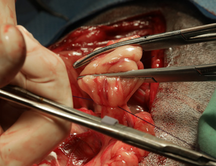
The final blood vessel to the spleen is cut while Jessica holds on to the spleen

After almost 2 hours of surgery Colt is now lighter on his feet

It’s official, at 14.095 pounds it is the largest spleen ever removed at Long Beach Animal Hospital

The vertical and long structure on the left is an enlarged and normal spleen, with the hematoma of this spleen on the right
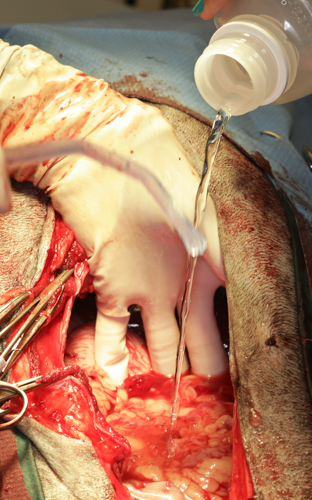
Surgery is nowhere near done. After Dr. K looks for any significant bleeding on the blood vessels she ligated, she flushes the abdomen numerous times with warm saline.
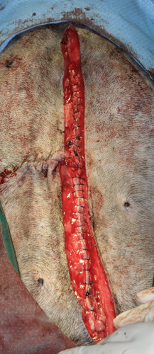
The long process of putting Colt back together now begins. The most important suture layer is an area called the linea alba. This is where the muscle bellies of the abdomen meet, and is a tendinous area that is very strong. This is where the first and most important layer of sutures is placed.

After more layers of sutures are placed, and before Dr. K puts in skin staples, Colt now has a bikini scar (OK, maybe not, but once the hair grows back nobody will know his secret)
In addition to the pain patch put on the night before, and the pain injection given during surgery, a long acting local anesthetic is placed at the incision. Now Colt will be comfortable when he wakes up and through the night.

All of our abdominal surgeries receive therapy laser treatment on their incisions. This aids in both healing and pain control. Notice the staples in Colt’s skin incision.
Once we have finished suturing our patient, who is already on a pain patch (Duragesic or Fentanyl patch- which is removed in 3 days), is given an additional pain injection and carefully monitored post-operatively.
As part of the monitoring we perform a simple blood panel to make sure there was no problem with blood loss during surgery. If the blood loss is significant we will give a blood transfusion with the blood we have already set aside specifically for this patient.

Now comes the bandage; the first layer is gauze and telfa pads
After more pads and gauze are put over the incision, the outer layer is wrapped by Terri while Jennifer and Jessica hold up Colt. They are getting their biceps exercise for the day!

We did the surgery the day after Halloween, so we thought this was an appropriate bandage

Our dedicated surgical team posing with their trophy. Great job!
Colt was closely monitored for several days, and his red blood cell count was checked to make sure he did not need that blood transfusion. He went home after 2 days and was back to normal in no time. Yea Colt!
Pathology Report

Great news on Colt’s histopathology report, confirming it is a hematoma. Colt should make a complete recovery and lead a normal, and lighter, life.

Just in case you want to see what a spleen hematoma looks like under the microscope here is your chance

Colt with his happy mom two weeks later when he came in for his staples removal
Case Summary
If you go back to Colt’s initial history, his mom stated that Colt vomited on occasion over the last month. Looking at the size of this hematoma (a record for Long Beach Animal Hospital), the hematoma was probably brewing over several months. It’s a wonder Colt could even eat, and was not vomiting more, with a hematoma of this size in his abdomen.
If Colt had not been brought in for his Wellness Exam, this hematoma could have easily ruptured, causing Colt to go into shock. and most likely a rapid death. Colt’s weight was the same as a previous visit. It was only because he had such a large mass in his abdomen, and goes to show you cannot go by just one physical parameter to determine health. It also shows you need to be thorough when investigating a medical problem.
The ability to do ultrasound with our highly skilled radiologist, telling us the hematoma was not actively bleeding, allowed us to take the time to prepare Colt for surgery. This made him a much better anesthetic risk, and and allowed him to heal faster with no complications.The ultrasound told us it was probably a hematoma, and there was no evidence of spread to the heart which would indicate a potential malignant hemangiosarcoma of the spleen. With the blood panel and chest radiographs being normal, it was realistic to proceed with surgery based on the fact that the mass was probably not a malignant cancer.
Now that you have seen a successful hematoma surgery, now might be a good time to learn about hemangiosarcoma, a malignant cancer of the spleen. In this same page you can see another hematoma surgery of a dog with an 8 pound hematoma of the spleen.