
A thorough approach is needed for a correct diagnosis of any liver problem. An organ like the liver that is so intimately involved with other important organs will exhibit symptoms that mimic disease in these other organs. Also, what initially might appear as a diseased liver is in reality a disease elsewhere in the body that is involved with the liver secondarily. This is why it is crucial to follow a thorough and methodical approach called the diagnostic process.
Signalment
Liver disease can occur in pets of any age. If it occurs in young animals we tend to think more of toxicity, a liver shunt or a viral disease like adenovirus in dogs, or FIP in cats. In older pets we tend to think more of inflammation and cancer, in addition to cholangiohepatitis and gall bladder problems, as the cause of the liver problem.
Several canine breeds are prone to getting liver disease:
Bedlington terrier’s, Skye terriers, Doberman pinschers, and West Highland White terriers get a problem with excessive copper accumulation that results from failure of normal biliary excretion of copper.
Cocker spaniels have an increased incidence of chronic hepatitis.
History
Early signs of liver disease are subtle, and might exhibit as some of the symptoms described above. It is important to remember that some pets do not show any symptoms early in the course of the disease. This is another reason for yearly exams, along with blood and urine samples in dogs and cats 8 years of age or more. Even though many cancers do not show up in a blood sample, we can sometimes get indirect evidence there is a problem, leading to additional diagnostic tests that might find cancer.
The recent use of pesticides, insecticides, and drugs might give us a clue. A history of poorly controlled diabetes mellitus might also clue us in to liver problems. Pets with liver shunts might have stunted growth and become depressed right after eating. In cats with hepatic lipidosis the history usually involves a lack of appetite (anorexia), especially if the cat was previously obese.
Symptoms of liver disease are variable and subtle in the early stages of the problem. The classic symptoms are:
Poor appetite (anorexia)
This is a common symptom and one of the primary reasons pets are brought to us for an exam.
Weight loss
The poor appetite that occurs in liver disease eventually leads to loss of weight. Improper metabolism of fat, carbohydrates, and proteins complicates the situation also.
Polyuria/polydypsia (PU/PD)
This is excess urinating and excess drinking of water. This can occur in liver disease, although several other important diseases cause these symptoms also, notably, kidney disease, Cushing’s disease, pyometra, and diabetes mellitus (sugar diabetes).
Lethargy
Poor appetite and disruption in normal physiologic processes leads to this symptom. Anemia adds to this lethargy, along with ascites due to the discomfort it causes.
Anemia
Improper nutrition from a poor appetite, along with disease in the hepatocytes will cause this.
Light colored stool
If the biliary tree is prevented from secreting normal bile pigments into the intestine the stool will lack pigmentation and appear lighter in color.
Bleeding disorders
The normal clotting system is impaired since it depends on a healthy liver.
Distended abdomen due to ascites or hepatomegaly. If the distention is severe enough breathing might be labored from pain or the pressure on the diaphragm.
Vomiting (emesis), nausea, or diarrhea
Sometimes blood is present in the vomitus (hematemesis), especially if a gastric ulcer is present. The ulcer comes from a complex interaction of histamine, nitrogen, bile acids, Gastrin, portal hypertension, and an altered mucous membrane lining the inside of the stomach.
Pain
Due to distention of a diseased liver.
Orange colored urine
Yellow mucous membranes
This is known as icterus or jaundice. It is a yellow discoloration of any part of the body that is white. It can also be caused from a severe anemia, so it is not always liver disease that can cause this.

This dog has a subtle icterus. It can be seen as the yellowish tinge to the white part of the eye (the sclera)
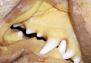
The icterus in the mucous membranes (gums) of this dog is obvious
Behavioral changes-
Circling, head tilt, heap pressing, and seizures, particularly right after a meal. These might be due to excessive ammonia buildup, seen often in a liver shunt.
In a recent study it was found that dogs with liver disease can also have high blood pressure. This is called hypertension, and should be monitored to see if therapy is needed.
Physical Exam

A physical exam on our patients is one of the most important ways to make a diagnosis in any disease
Distended abdomen due to enlargement of the liver (hepatomegaly) might be found. This can be palpated in some situations, especially in the smaller animals. an enlarged liver from a disease other than liver disease can cause hepatomegaly. This includes heart disease and Cushing’s Disease.
Enlarged lymph nodes due to secondary bacterial infections or spread of a primary or metastatic liver tumor.

This cute Sheltie is having its submandibular lymph nodes palpated as part of our routine exam
Bruising (hematoma) might be observed under the skin, or when a blood sample is obtained. This is due to the liver’s affects on the clotting mechanism.
Fever- a rectal temperature of greater than 103 degrees F could accompany liver disease when inflammation or infection is present.
Skin infections and wounds that do not heal, or recur after antibiotics are stopped.
Anemia might be observed by checking the mucous membranes for a normal pink color.
Diagnostic Tests
Several tests are used as an aid in making this diagnosis.
Blood Panel
A CBC (complete blood count) and BCP (biochemistry panel) should be run on every pet 8 years of age or more, especially if they have any of the symptoms of liver disease.

The CBC might show a decrease in the number of red blood cells (RBC’s). This decrease in RBC’s is called anemia. The white blood cell count (WBC) might be elevated (leukocytosis), normal, or decreased (leukopenia), mostly depending on the cause of the liver problem and how long it has been present. A change in the WBC’s does not necessarily indicate there is a liver problem.

The biochem panel (BCP) might show an elevation in the liver enzymes. If a dog or cat has normal live enzymes on the blood panel, it still could be liver disease. This is because the liver cells are so scarred down (cirrhosis) that there are no enzymes in them to leak out into the bloodstream and detection by our blood panel.
Bile Acids
This is liver function test, not an enzyme test, and is not a routine part of the BCP. We will request this test when we suspect a liver problem, whether the enzyme tests are normal or not. This test is performed by taking a blood sample, giving a meal, then taking another blood sample 2 hours after the meal. Comparing the pre-meal and post-meal blood results gives us valuable information. The bile acids test is an accurate measure of liver function.

This one is very high. This pet has a liver shunt
Urinalysis
A urine sample can give us important clues as to the existence of liver disease. The specific gravity might be below normal, an indication that PU/PD is present. Bilirubin might be present, a finding that is always abnormal in cats. There also might be ammonium biurate crystals, a sign of improper ammonia metabolism found in Hepatic Encephalopathy.

This spillover of bilirubin from the bloodstream to the urine is a sign of liver disease, especially in the cat
Abdominocentesis
Analysis of the fluid obtained from a pet with ascites can give valuable clues as to its cause. There are numerous causes to ascites, some of the more common ones are heart disease, liver disease, and cancer.
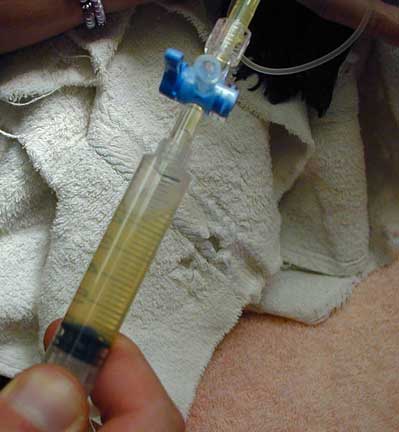
Fluid is removed from the abdomen with a special needle and syringe
Liver Biopsy
This is a very valuable test in the diagnosis of liver disease. A sample of the liver can be obtained during an exploratory surgery or during an ultrasound procedure. The pathologist can look at the hepatocytes microscopically and determine if disease is present and what the cause is.

This is how we take a liver biopsy during an exploratory surgery
This report is from a very ill cat.

It is helpful to run a coagulation panel prior to any liver biopsy. A diseased liver might not be able to clot properly, and a biopsy could cause hemorrhage into the abdomen.

This Coagulation Panel is normal, so we are OK to biopsy this liver and not worried about hemorrhage
Stool
A dog that excretes stool without normal pigmentation could indicate liver disease. It occurs when there is obstruction of the biliary system and normal bile pigments are not secreted to cause the normal dark color of stool.
Radiography
An enlarged liver on a radiograph is called hepatomegaly, an abnormally small one is called microhepatica. Either one can be a sign of a liver problem.
In addition to plain radiographs, contrast media can be put into the arterial or venous system to help outline the liver. These tests go by various names; cholecystography, portal venography, and hepatic arteriography.

The liver in this radiograph is enlarged because the edge of the liver is protruding far beyond the last rib. The edges of this liver are very sharp and clearly outline its borders.

This radiograph also shows hepatomegaly, but in this case the borders of the liver are not as sharp. This could be due to a swelling of one of the lobes or fluid in the abdomen. An enlarged spleen can look like this also.

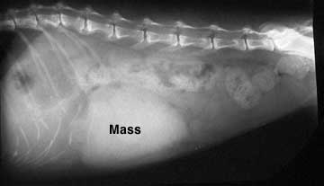
Some radiographs of a liver with hepatomegaly don’t show the routine shape of the liver lobes. This case of a liver cancer has a very rounded appearance. A tumor of the stomach, spleen, or intestines can also have this appearance.

Sometimes we diagnose hepatomegaly or microhepatica indirectly by looking at the angle of the stomach This picture shows the angle of the stomach in a normal radiograph of the abdomen. Compare it to the radiograph below.

This enlarged liver is pushing the stomach (S) towards the rear, an indication of hepatomegaly, even though it is difficult to clearly see the liver.
Sometimes we can not say for sure whether an enlarged organ on a radiograph is the liver. This mass, located near the liver, could also be an enlarged spleen, small intestine, lymph node, stomach, or even pancreas.
Ultrasound
Ultrasound is highly beneficial in the diagnosis of liver disease. We recommend ultrasounding a liver when the liver enzymes tests are elevated over time, or the bile acids test is abnormal. Ultrasound has replaced exploratory surgery in many cases because it is less expensive, far less invasive (all we need to do is shave the tummy) and highly accurate when performed by our radiologist Ann Reed.

You can easily see a lobe of the liver in the center of this abdominal ultrasound of a dog
The internal structure (called parenchyma) can be analyzed, and post-hepatic liver disease can be differentiated from hepatic liver disease. This can be very important because disease in the liver can often be diagnosed with a biopsy during the ultrasound. Post-hepatic liver disease cannot easily be diagnosed in this matter. Instead it is diagnosed and treated with an exploratory surgery (called a laparotomy).

This liver ultrasound reveals a mass in the liver. Can you see its circular appearance at the arrow? It also shows abdominal effusion (this is the ascites described previously).
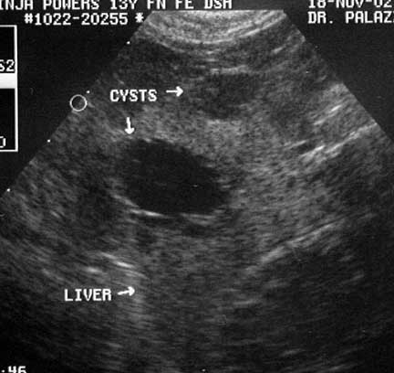
This is what liver cysts look like on ultrasound

This is the detailed ultrasound report we get when the liver and other abdominal organs are analyzed

The gallbladder, which lies in-between liver lobes, is circled

When there is a serious problem with the gallbladder it needs to be removed. This requires the expertise of an experienced surgeon because the gall bladder is so closely associated with the liver.

Our surgeon is using a sterile Q-tip to gently dissect the gallbladder away from a lobe of the liver. Click here to see the full surgery.
Continue on to routine medical treatment of specific liver diseases by clicking here.