An important diagnostic modality we use to make an accurate diagnosis on our dog patients is radiography, more commonly known as x-rays. Just like the many other Diagnostic tests we perform, x-rays are important in veterinary medicine since our patients do not routinely tell us where they are having a medical problem.
We work closely with our radiologist Dr. Ann Reed, along with imaging specialists from Antech Imaging Services (AIS) to aid us in using X-rays for diagnostic purposes.
This fun section shows you the wide variety of radiographs we take on dogs. On many of the radiographs there is a link to the canine disease process that is going on for much more information.
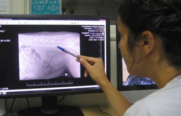
It takes many years of experience to learn how to read a radiograph. This is one of our student externs honing her skills even before she gets out of veterinary school.
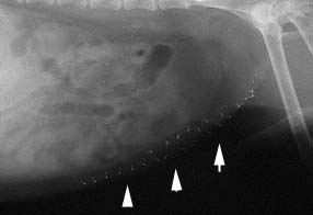
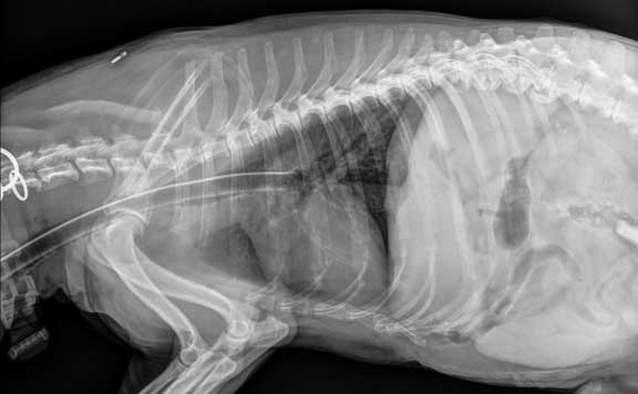
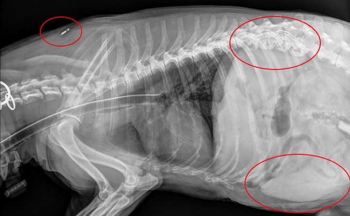
All of these metallic objects (see arrows) are stainless steel sutures in Spunky. He is a Schnauzer dog that has gone through 3 abdominal surgeries. Two have been to remove bladder stones, one was to remove something he ate (you wouldn’t want to know what it was) that got lodged in his stomach.
The round white thing on the far right is a bladder stone (urolithiasis) in a canine. Click here to learn all about these stones.
The diffuse white area in the center of this dog’s abdomen is an abdominal tumor. It most likely is a splenic hematoma , or it could be a tumor of the spleen called hemangiosarcoma. Click here to find out and see how we took care of it.
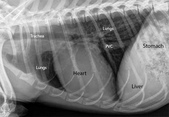
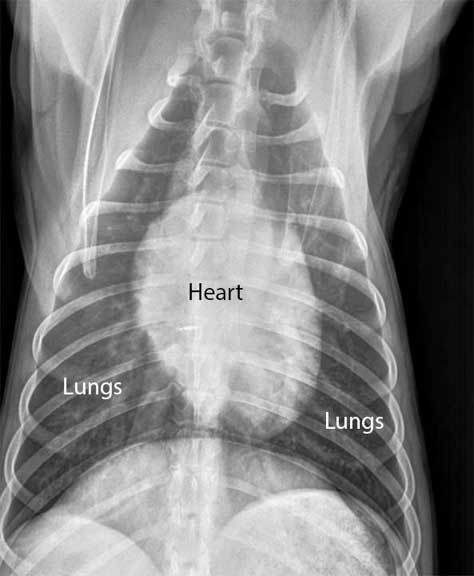
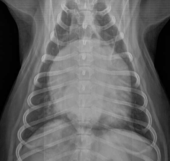
The following radiographs are of a dog’s chest. The first two normal ones are labeled so that you can see the problem in the two with the enlarged heart. An enlarged heart is called cardiomegaly, and can be due to heart failure.
Lateral view of a normal chest. PCV is posterior vena cava.
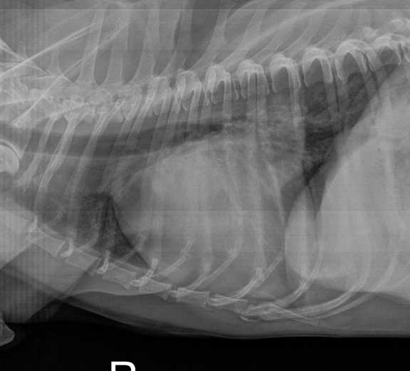
Notice how large the heart is and how the trachea is being pushed up by this enlarged heart
DV (dorsoventral) view of the same dog
Look at how much more space the enlarged heart takes up in this radiograph of the same dog
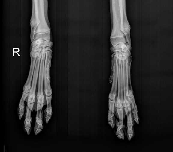
Lots of bones in the front legs and feet of a dog:
Radius
Ulna
Carpals
Metacarpals
Phalanges
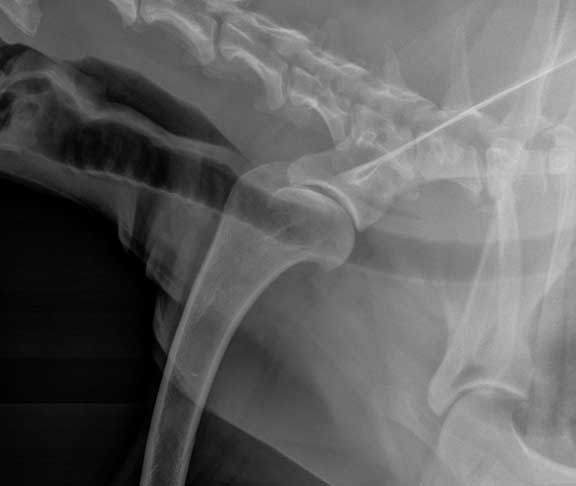
The shoulder joint, with the scapula and humerus also. The top left horizontal black area is air in the esophagus. The bottom and much longer horizontal black tube is the trachea (windpipe).
X-Ray of the breathing tube used to give oxygen and anesthesia during surgery. It is the horizontal white tube from the left, and it is in the trachea. Notice anything else on this radiograph?
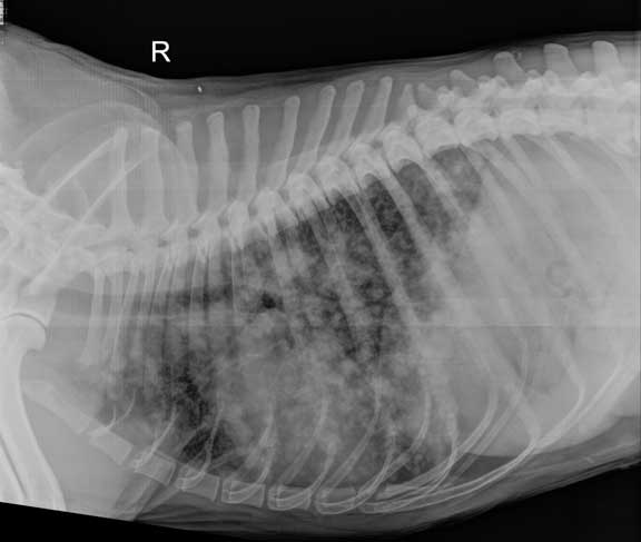
This chest is filled with cancer. It is that moth-eaten appearance.
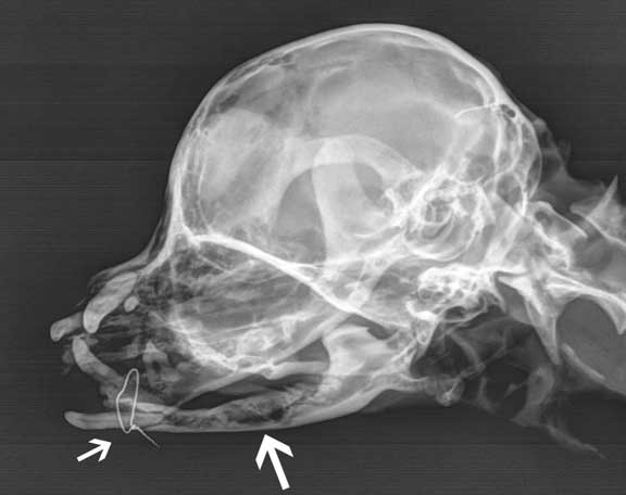
The wire is in this mandible (lower jaw) to hold it together because it spontaneously fractured due to advanced dental disease. The larger area points to the diseased jaw. Severe dental disease is common, so please read our Dental Page to learn how to prevent this serious problem in your dog.
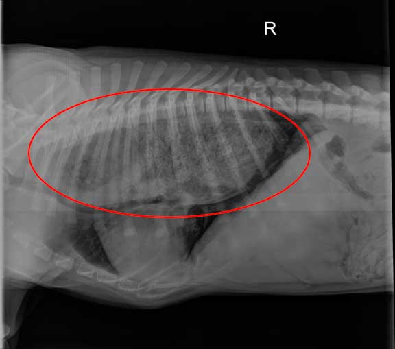
The circle in this dog’s chest radiograph is over the esophagus that is filled with food. This is called megaesophagus. In this disease food does not readily pass down the esophagus into the stomach, and pools partway down.
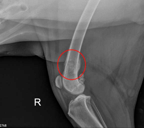
The fuzziness on the cortex of the bone, and the dark area (called lysis) in the shaft of the bone is a cancer called osteosarcoma.
The last radiograph goes back many decades when Dr. P first started working at Long Beach Animal Hospital in 1989. It is from a dog that came in straining to defecate as if it was constipated. The dog had been given several different treatments for other veterinarians for his constipation to no avail.
Dr. P took a radiograph and found something every interesting in the rectum. It was a stack of pennies!