
Introduction
This is a fun and educational page for pet owners, veterinary students, technicians, and even other veterinarians. It is designed to educate you on the basics of radiology. There is a fun test at the end to see how much you learned. Hopefully you will get them all right!

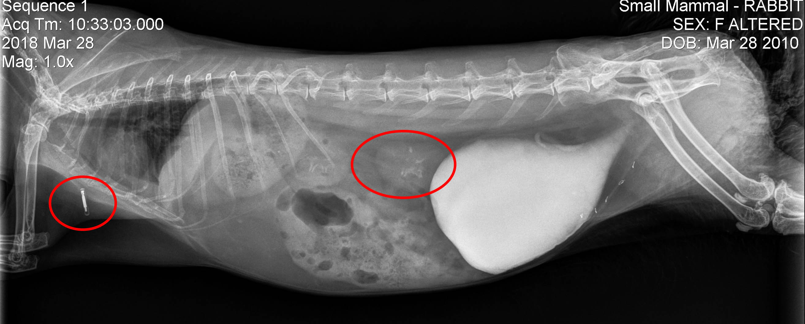
Can you tell what animal this is from this radiograph? Hint: it is a reptile
An important diagnostic modality we use to make an accurate diagnosis on a sick animal is radiography, more commonly known as x-rays. Just like the many other Diagnostic tests we perform, radiograph x-rays are important in veterinary medicine since our patients do not routinely tell us where they are having a medical problem.
Learning how to accurately read a radiograph on the wide variety of species that we care for requires requires a large body of knowledge. It is an ongoing process for our veterinarians in order to develop this skill. We oftentimes utilize the expertise of a veterinary specialist in reading radiographs of animals in the more complicated cases.
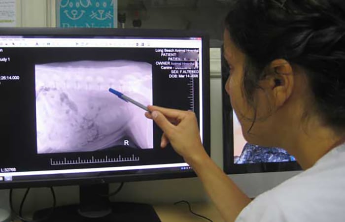
With such variability on what is normal or abnormal, many radiographs of the non dog and cat (we call them exotics) pets that we care for at the Long Beach Animal Hospital require the assistance of a specialist. The lab we use for our blood panels has a division called Antech Imaging Services (AIS) that gives us a detailed and written interpretation of these radiographs within 24 hours on all species, including the exotics. They have a veterinarian that is board certified in zoo animals to help us in interpreting the more difficult cases in exotic species.
The veterinarian that reads radiographs for AIS is Marie Rush. Marie is dedicated to her profession and the well-being on sick animals. Even when she is out of the country doing conservation work she is still able to provide a turnaround time of 2 hours on interpretation of radiographs that are sent to her, although she has different set of office furniture while in Costa Rica.
We start our students down the path of this lifetime commitment to learning this skill early in their careers when they join our student externship program
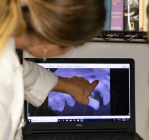
Our digital radiography has dramatically enhanced our ability to find problems, and is especially useful when we do dental work on pets
We work closely with our radiologist Dr. Ann Reed, along with imaging specialists from Antech Imaging Services (AIS) to aid us in using X-rays for diagnostic purposes.
Before we get started reading radiographs, let’s get some basics out of the way.
Views
Several different angles are used to assess radiographs. The two that are used the overwhelming majority of time are:
- the lateral (side) view where a pet is laying on its right or left side
- the ventrodorsal (VD) view where the pet is laying on its back. There is also a DV view with a pet laying on its abdomen
In this lateral view of thi fat cat the “R” means it is laying on its right side
The same fat cat in this VD view. The “R” marker shows the right side of the cat
Radiographic Densities
There are five radiographic densities:
Soft tissue– internal organs like the liver and kidneys that have a whitish color
Fat– the fat around the internal organs, also with a whitish color. Without this fat you would not be able to differentiate the different internal organs like the liver or kidneys, since they are soft tissue, and have the same radiographic density.
Air– this is black, and is what you see for the lungs in a chest radiograph
Bone– which is brighter than soft tissue or fat
Metal– Vivid, very bright, and hard to miss
Interpretation
Look at each x-ray closely (sometimes very closely) and see if you can figure out what is wrong. We have a couple of clues to help you make an interpretation:
- Use symmetry when you can. Compare both sides, legs, or whatever else that might be useful.
- Pull your face away from the screen and scan the whole x-ray before you jump into the details.
- After you have scanned the whole radiograph look very closely for subtle changes.
First we will show a bunch of fun radiographs of the more unusual pets we see at our hospital. After that we will do some radiograph reading lessons, teaching you about the normal anatomy of dogs and cats. After that is a little test to see how you did. We will stick to abdominal radiographs for the test to make it easier. Good luck, and have fun!
Exotic Animal Radiographs
These first few rads are helpful to get your eyeballs warmed up for your test at the end.
Pregnant Guinea Pig. How many piglets do you see?
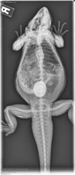
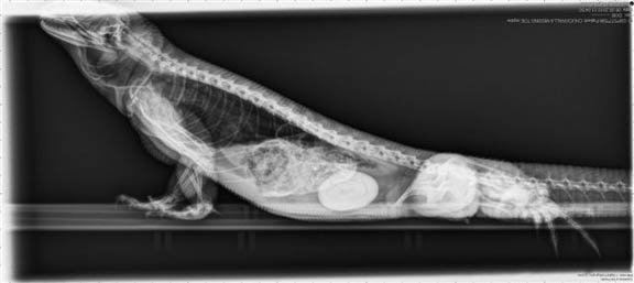
Iguana bladder stones. Click here to see the surgery to remove a bladder stone in an Iggie.
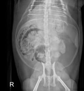
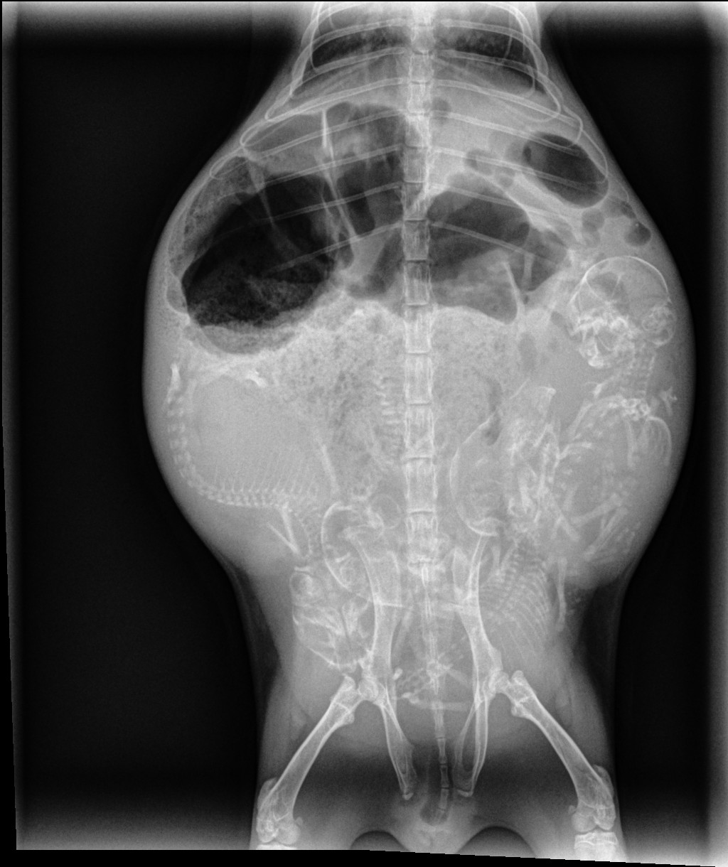
Female rabbit with mummified fetuses that are several months old
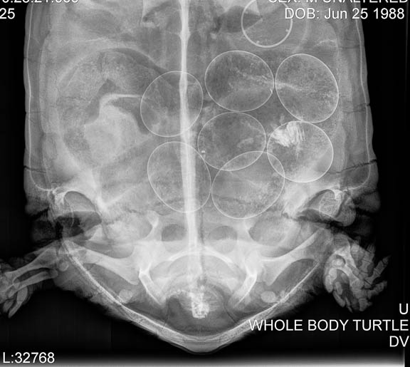
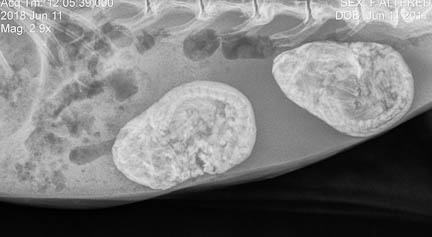
California Desert Tortoise (CDT) with eggs. Click here to see what they look like inside of a different tortoise when we do surgery to remove bladder stones.
Two small and white bladder stones in a Guinea Pig

Normal hawk from our Wildlife Care Program
Hawk with a broken wing (technically a mid-shaft fracture of the humerus) from our Wildlife Care Program
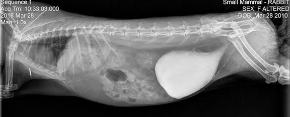
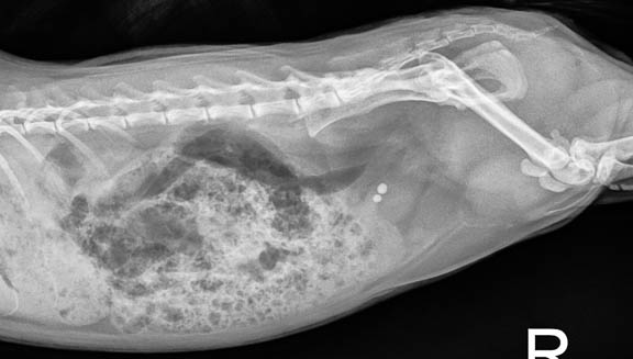
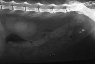
Calcium sludge in the bladder of a rabbit. This is called hypercalciuria, and you can read our detailed page on it.
Did you also see the microchip and the calcium in the kidney?
Chinchilla incisor and molar teeth
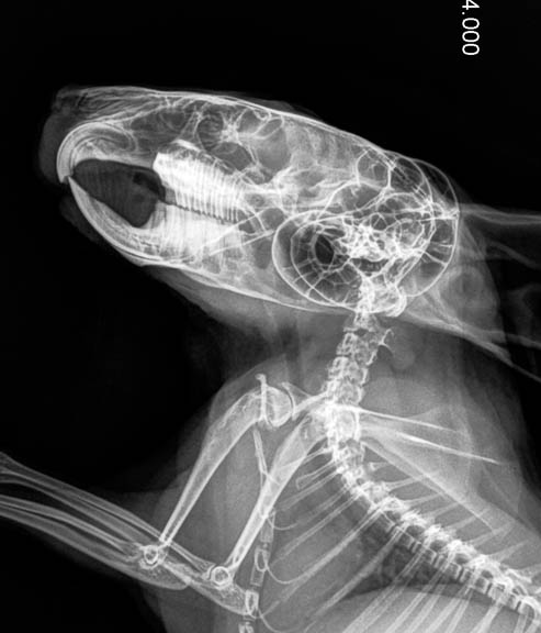
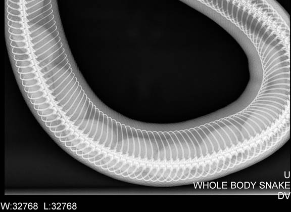 Snake with eggs

Rabbit with a fluid filled uterus. To see how we took care of this problem follow this link.
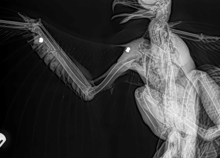
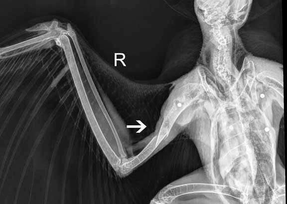
Do you see the two pellets in this hawk’s wing?

Did you also see the fracture circled in red at the tip of the wing? How should this be handled? You can see what we did in our Wildlife Care Page
Normal X-Rays of dogs and cats
Enough of these exotics and wildlife, let’s get to the dogs and cats. They are mammals like us, and are easier (we didn’t say easy, though) to interpret than the exotics.
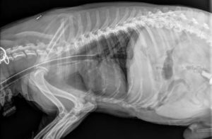
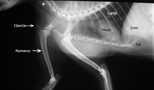
A lateral X-ray of a dog’s chest and cranial abdomen. The head is at the far left.
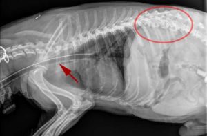
Same X-ray as above, with an arrow pointing to the breathing tube for anesthesia, and the arthritis in the spine, circled in red
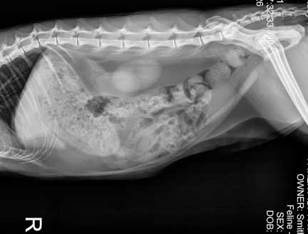
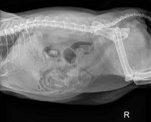
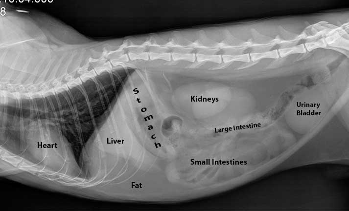
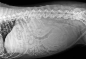
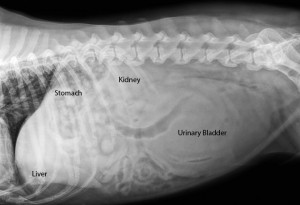
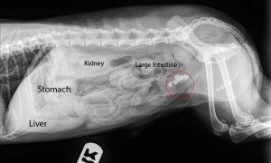
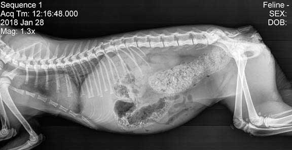
This is a radiograph of the abdomen of a normal cat that is laying on its right side. The head is towards the left. Use the diagram below to identify the organs.
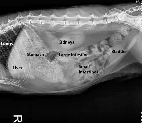
The stomach has food in it, and the large intestine contains feces. All five radiographic densities are present in this abdominal radiograph. Do you see all of them?
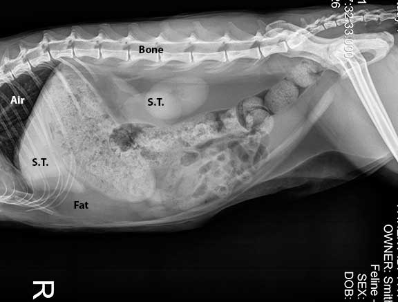
Air- is in the lungs along with gas in the intestines
S.T. -soft tissue is the liver and kidney
Fat- this is abdominal fat, also known as falciform fat
Bone- lumbar vertebrae
Metal- the R marker to indicate this cat is laying on its right side is made of metal
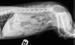
Here is another normal cat abdominal and chest radiograph, this time with an empty stomach
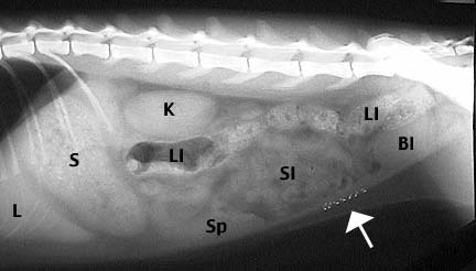
Here is another one, this time with the spleen and metallic sutures from a spay. You can easily see the liver (L), stomach filled with food (S) kidneys (K) , the small intestines (SI), the large intestine (LI), the urinary bladder (Bl), and the Spleen (Sp). The arrow points to stainless steel sutures in the muscle layer from a spay operation.
Abnormal X-Rays
Now that you know how to read normal dog and cat rads let’s look at some abnormal ones.
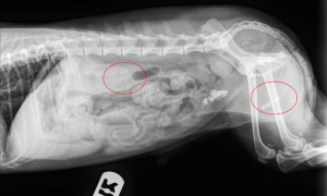
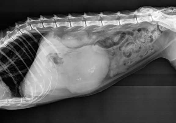
This dog is having a difficult time urinating. Can you tell what is wrong?
Look towards the right side of this abdominal radiograph
Does labeling the organs help in your diagnosis?
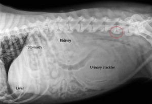
The bladder is huge, because this dog is having a difficult time urinating. It is probably due to nerve dysfunction, since the spinal cord has changes called spondylosis. The circle points this out on one of the vertebrae
You can learn more about this problem, called spondylosis, from our arthritis page
This is a dog abdominal radiograph. Notice anything unusual?
Again, look towards the right side
You can see the circle around the numerous stones (called calculi) in the urinary bladder
Did you also notice the stones in the kidney and pelvic urethra?
Our web page on bladder stones has lots of good information on how we diagnose, treat, and prevent recurrence, of this disease.
This cat is labeled for you. Anything fishy?
Look towards the left side of the radiograph this time
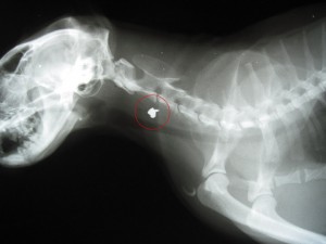
Did you see the pellet in the neck? Look again at the radiograph above, it’s plain as day.
Now that you are an expert at reading radiographs give the following one a try. It is from a cat that is straining to urinate and has blood in its urine. The answer is below, along with a picture with arrows pointing to the abnormalities.
This cat has 2 stones in its urinary bladder (click here to learn more about them and see a surgery of how they are removed). The stones are radiopaque, which means they show up easily on the radiograph. Some bladder stones are radiolucent, and can only be seen by injecting dye or air into the urinary bladder.

The arrows point to the bladder stones, along with the faint metallic sutures from a spay
Pretty easy so far, huh? Don’t get too confident just yet, our next few are a little harder. Look over the next few abnormal radiographs and send us an e-mail with your answer. If you aren’t sure and just need some clues e-mail us also and we will help you. Good Luck!
Abnormal X-Rays
Now that you are experts at reading x-rays, you can put your newfound skills to work. Email us at vet@lbah.com for the answers.
- What do you think about this cat radiograph?
2. This radiograph is an abdomen view from a very sick dog. It is 13 years old and losing weight
3. This is from an elderly dog that is losing weight

3. This dog is limping on its rear leg
