
Laser Energy
The word laser stands for Light Amplification by Stimulated Emission of Radiation. In a carbon dioxide laser an electrical current that changes from positive to negative 40 million times each second excites a carbon dioxide molecule to a higher energy state. This carbon dioxide molecule then sheds this higher energy as a photon, or wavelet of light. As soon as the carbon dioxide molecule sheds this energy as light it is available for re-excitation.
Some of the photons emitted are emitted along the axis of the laser tube. They bounce back and forth between mirrors that are at each end of the laser tube. When an excited carbon dioxide molecule is struck by a photon flying between mirrors it is stimulated to emit a photon that is identical to the stimulating photon. This is the Stimulated Emission. The original and newly admitted photons continue to stimulate the production of more photons resulting in an amplification of Light. Some of these photons are allowed to escape through a special transmissive mirror. Those that escape create the laser beam.

Laser Classes
-
Class I
Grocery store scanners are in this class. They are no more harmful to the eye than an electric light bulb.
-
Class II
Laser pointers are in this class. Momentary viewing of these lasers is not considered dangerous.
-
Class III
Laser Light Show. These lasers may be hazardous when viewed directly.
-
Class IV
Most surgical lasers are in this class. They can cause serious eye and skin injury and set fire to many materials. Both the direct and reflected beams are hazardous to the eye.
Wavelength and Tissue Type
Lasers of different wavelengths produce different effects on tissues. CO2, Nd:YAG, diode, and argon lasers cause tissue to heat up. The wavelength of the laser determines the absorption into various structures such as h20, melanin, and hemoglobin, etc. Several things happen in general when light from these these types of lasers strikes tissue:
-
The top layer of tissue heats up and is vaporized.
-
Beyond the cut, a narrow zone of tissue is rendered necrotic by the beam and by heat conducted from the vaporization zone. The disadvantage of this necrosis is that tissue is damaged, the advantage is the control of small blood vessels at the edge of the cut.
-
Beyond the zone of tissue necrosis the tissue is affected by heat but is potentially still viable.
The CO2 Laser
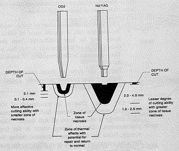
Due to its wavelength of 10.6 micrometers, CO2 laser energy is efficiently absorbed by water. Soft tissue is greater than 90% water, so it is effective on all soft tissue, whether the tissue is pigmented or not. Because of decreased water content in bones and teeth this laser is not used on these structures. During lasing, water in the target tissue absorbs the laser energy, heats up, boils, and vaporizes, taking the surrounding tissue with it. Because energy absorption is so efficient with the CO2 laser on soft tissue that energy penetrates no more than 0.1mm beyond the cut, regardless of the cut depth. The zone of necrosis and thermal injury under the cut is only 0.5 mm in depth. This allows precise control by the surgeon.
The Nd:YAG Laser / Diode
With a wavelength of 1064 nM, Nd:YAG laser energy is less efficiently absorbed by water and more readily absorbed by other materials such as the hemoglobin in red blood cells. The energy penetrates further into the soft tissue (up to 4 mm) before being absorbed. Because the energy is diffused into a larger volume of tissue compared to the CO2 laser, cutting is less efficient and the zones of necrosis and thermal injury are larger. This may be beneficial when removing a tumor deep in the body. By intentionally cooking it the tumor is destroyed. The absorption efficiency is dependent on the pigmentation of the tissue.
The real advantage of these wavelengths is that they are delivered through a glass fiber, therefore they can be passed through an endoscope. Even though they can’t cut as good as a CO2 laser they are able reach areas the CO2 laser can’t.
The relative effect of the CO2 and Nd:YAG lasers on tissue. Note the large depth of cut and smaller zone of necrosis of the CO2 laser compared to the Nd:YAG. More necrosis means better control of bleeding, but too much increases healing time. The CO2 laser provides a nice balance for general incisional work.
Power and Exposure Time
Power Density
This is the power delivered per unit area, expressed usually as watts per square centimeter. Power density depends on the set power, the spot size, and the distance of the tip from tissue.
Spot size
The spot size is the diameter (in mm) of the aperture containing 86% of the total power of the laser beam. If the power is held constant, a larger spot size produces s smaller relative impact on tissue than a smaller spot size, because the power is spread over a larger area.
There are two factors which determine the spot size of the hollow waveguide delivered CO2 laser on target tissue:
Rated spot size for the tip in use
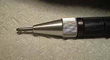
This is what a 0.4 mm tip looks like at when attached to the hand piece.
Distance of the tip from the tissue target
This diagram shows the effect of tip size and target distance on tissue spot size.

Program and Exposure Options
In the LX-20LP laser power is generated as a continuous wave:
-
In continuous exposure, lasting as long as the foot switch is depressed
-
In single exposure one time exposure is delivered for each depression of the foot switch
-
In repeat exposure the laser beam cycles between on and off while the foot switch is depressed
The arrow points to the three different exposure settings on the machine. The top is single exposure, the middle is repeat, and the bottom is continuous. This machine is set at 6 watts of power and 40% program (it is ON 40% of the time).

Incision
To cut or incise tissue focus the beam and use the tip at its nominally rated spot size ( 1/2 to 3 mm from the target tissue). Power and time determine the depth of cut. In general, use 6 watts of continuous exposure, or for better control, repeat exposure with a high duty cycle (40% program). Use a 0.25 or 0.4 mm spot size tip.
Excision
To excise, focus the beam ( keep tip 1-3 mm from tissue) and repeat exposure. Use a 0.4 mm spot size tip, then excise using 12 watts (20% program).
Ablation
To vaporize you can use a focused or defocused beam depending on the amount of tissue to be removed and the precision required. In general, use a low power setting (4-10 watts) and continuous exposure, or a higher power setting in repeat exposure. Ablate the tissue layer by layer with a sweeping motion and wipe away the char after each layer. Char absorbs CO2 laser energy and will overheat the underlying tissue. It is possible to sculpt contours, an advantage over the scalpel.
Hemostasis
For hemostasis use 3 watts continuous exposure, defocused (pull the tip back 4-12 mm from target tissue). Use a 0.8 mm spot size tip and lase the vessel, not the pool of blood. Lasing is effective on vessels that are less than 0.5 mm diameter.
Laser Safety
A laser is a high energy beam of infrared radiation in the form of light waves that poses potential health hazards to patients, operators, and bystanders. It is imperative that safety procedures are followed. Wet gauze provides an effective safety backstop for CO2 lasers because the energy is absorbed into the water.
Hazards associated With Laser Use
- Eye hazards
-
Class IV lasers are very damaging to eyes. Everyone in the immediate vicinity of the patient must wear protective goggles rated for a CO2 laser. The patient should have protective covering over the eyes.
- Skin Hazards
-
The skin is the second most vulnerable organ in the body to lasers. Direct and reflected laser light can cause thermal injury to the skin. Drapes and wet packs should be used for the patient.
- Smoke plume hazards
-
The interaction of laser with tissue produces a plume of smoke that may contain toxic and carcinogenic chemicals. Viruses might also be present. The use of 0.2 micron masks or smaller and smoke evacuators is mandatory
- Fire hazards
-
Drying agents, ointments, plastic resins and anesthetics can also cause a fire when exposed to a laser. Alcohol preparation of the skin prior to surgery is also a fire hazard and should not be used. Surgical drapes are flammable when exposed to a laser and should be kept wet with saline at all times. Endotracheal tubes are a serious hazard potential due to their flammable nature and the fact that they are filled with oxygen. They should be packed off with saline soaked gauzes or special endotracheal tubes should be used. Oxygen use in general is hazardous along with flammable anesthetics and methane gas from the lower digestive tract. The rectum should be packed with sterile saline whenever anal surgery is performed. always have a fire extinguisher nearby.
- Gas embolisms
-
The laser unit emits purge gas under pressure when lasing. To reduce the risk of gas embolism do not bring the tip into contact with any vascular tissue.
- Electrical and facility hazards
-
The interior of the laser unit contains exposed high voltages and invisible laser light. Internal maintenance is to be performed only by trained technicians.
Daily Checklist
Before the daily use of the laser a routine checklist should be followed:
Warning notice posted.

Only personnel trained in laser safety are present.

Safety goggles in use.

0.1 micron masks used

Patient eye protection in place.

Smoke evacuator operational

The LX-20 Laser
This is the operational end of our machine. You can see the on/off key at the top in the middle. The silver knob below it is the manual safety shutter. The red knob to the left of the safety shutter is the emergency shutoff switch.
On the main panel you can see the setting of 6 watts and 40% program. On the right side are the standby and ready switches.
