
The lymph nodes are part of the lymphatic system. They have several important functions regarding the immune system, and are good indicators of disease. The mammalian body has many internal lymph nodes. These can only be visualized by exploratory surgery, a CT scan, an MRI, a radiograph, or an ultrasound.
If you would like to compare animal to people lymph nodes the Mayo Clinic web site has a nice explanation. Check it out after reading this page.
During a physical exam the external ones can be palpated to determine if they are enlarged. The medical term for enlarged external lymph nodes is lymphadenopathy. The lymph nodes are always examined as part of our routine head to tail (if they have one!) exam on all pets presented to us.
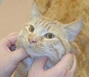
 Dr. P is getting ready to perform a lymph node exam on this cat. He will start with the paired lymph nodes under the jaw, called the submandibular lymph nodes.
Causes
Lymph nodes can enlarge due to several reasons:
-
Inflammation
A reaction to a foreign body might cause this. If mild this will go away by itself in most cases. If not, we will use medication like cortisone to get the swelling down. Even though this type of problem is benign, if the lymph nodes get large enough they can interfere with normal physiologic functions like swallowing.
-
Infection
Bacterial, viral, or fungal infections can cause them to enlarge. This enlargement might be a sign that they are performing their job as expected. When the infection subsides the lymph node should return to normal size. Sometimes though, an enlarged lymph is a sign that the lymph node needs help in fighting inflammation or infection, and we put that pet on antibiotics or anti-inflammatory medication.

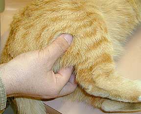
This badly infected toe on this cat could cause the regional lymph node, in this case it would be the axillary lymph node, to be enlarged.
-
Cancer
The most common cancer that causes this problem is called lymphoma (also know as lymphosarcoma). This is malignant cancer.

This is the report of a dog that has lymphoma
Examination
Internal Lymph Nodes
Numerous lymph nodes exist within the thoracic and abdominal cavities. They cannot be palpated, and their enlargement does not show up on routine blood samples. They might be seen on radiographs or ultrasound if they are significantly enlarged.
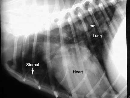
This radiograph of the chest shows the heart and lungs clearly. The lower arrow points to the location of the sternal lymph node. It is barely visible in this dog because it is normal. The top arrow points to a round, white structure. It is a tumor nodule that has spread from cancer elsewhere in body.

This radiograph of the abdomen shows the liver and stomach clearly. The middle arrow points to the general location of the mesenteric lymph node. The top right arrow points to the location of the sublumbar lymph node. Neither one is visible because they are normal.

The greatly enlarged sublumbar lymph nodes are circled in this dog with cancer. See the close up below

The enlarged sublumbar lymph nodes are visible as the whitish areas under the red circle. This is from a dog with a cancer called adenocarcinoma.
Lymph nodes can also be seen with the ultrasound. This takes an experienced veterinarian, usually a veterinary radiologist.

This is a lymph node in the center of the abdomen called the mesenteric lymph node

This is a lymph node at the back of the abdomen just under the spine called the sub lumbar lymph node. It is the same lymph node as the dog above.

This is a close up of a lymph node when seen by ultrasound
Enlarged lymph nodes can be found when performing an exploratory surgery.

The scalpel blade is pointing to the mesenteric lymph node. This node is found at the center of the small intestine loops. This one is enlarged because this cat has kidney cancer, which has spread to the mesenteric node.

Here is another enlarged mesenteric lymph node from a cat with intestinal cancer called lymphosarcoma
External Lymph Nodes
Mammals have numerous external lymph nodes (also called peripheral lymph nodes) that can be palpated during a physical examination. This is of great advantage because we can easily feel if they are enlarged without having to rely on diagnostic tests.
Reptiles and birds do not have these external lymph nodes, so they cannot be palpated during an exam.

Good luck trying to palpate the external lymph nodes on this 120# python in for its yearly exam. The only thing you will find is how powerful it is as your assistants try to hold it still for an exam.
The tonsils are lymph nodes also, but they are not palpated, just looked at during our routine exam. We do not recommend that you check them yourself because they are deep in the oral cavity, and can be confused with the organs. Also, you can injury your pet while opening the mouth wide enough to see them, or you can be injured by a bite or scratch from your pet. If you think there is something wrong with your pet’s mouth it should be brought to us for a proper and thorough exam.
Five external lymph nodes are routinely palpated during a physical exam. All of them are paired, which is another advantage because we can compare one to another and notice subtle changes in size. We will teach you how to palpate them during our exam.
-
Submandibular

They are located in the neck area near the angle of the jaw. They lay right next to the salivary glands, so it is important to palpate the correct structure. -
Prescapular

They are located just in front of the shoulders. Follow the arm up to the point of the shoulder to find them. -
Axillary

Found on each side of the chest in the armpit area. They tend to be small, and are more forward then most people realize. You need to be under the arm to feel them against the rib. -
Inguinal

Found on the abdomen towards the inside of each rear leg. There is significant fat in this area, so they might be difficult to find, even if enlarged. Also, there is mammary tissue in the area that can easily feel like an enlarged lymph node if the mammary tissue has an infection or cancer. These are not lymph nodes you would routinely palpate on your own. -
Popliteal

Found on each rear leg on the opposite side of the knee. They are pea sized, and relatively superficial and easy to feel.


We will gladly teach you how to palpate these lymph nodes whenever you bring your pet in for a physical exam. These are sensitive organs so they are always palpated in a gentle manner. You might want to palpate them on a weekly basis at home. If you think they are enlarged bring your pet in for an exam so we can determine if there is a problem.
We have a page in our Learning Center that shows you how to do an in-home exam on your pet. This section goes over the lymph node exam again, this time on a cute white dog.
Diagnosis
Fine Needle aspiration
This test is a relatively simple way to obtain a significant amount of information from an enlarged lymph node. In this test we use a small needle (similar to the one we use to give vaccines) and gently insert it into the enlarged lymph node. We aspirate some of the lymph node material and put it on a microscope slide.

This ferret has a very large right submandibular lymph node. Do you see the needle on the left side of the picture that we are using to obtain the aspirate sample? It is super sharp, and is no more uncomfortable than getting a vaccine.

The aspirated material is put on a microscope slide for analysis by a pathologist. They will put special stains on the material and carefully scrutinize it under the microscope.
The fine needle aspirate test is not infallible at finding a problem. When the needle is inserted it is put in only by feel. It is possible for the lymph node to have a problem area that the needle missed. Also, we are giving the pathologist only a very small sample to read. It can be difficult to determine the health of the whole lymph node when only a small sample is taken.
Due to these limitations the pathologist sometimes cannot say for certain what caused the lymph node to enlarge. If it comes back inconclusive then we might keep the lymph node under close observation. If there are other changes in the rest of the physical exam, or laboratory data indicates there might be a significant problem, we will remove the whole lymph node and submit it for analysis.
If the fine needle aspirate report comes back that cancer is suspected we will remove the lymph node, no matter what the other laboratory tests indicate. You saw that earlier when we showed you the report of a dog with lymphoma.
Biopsy
The most accurate way to determine if a lymph node is seriously diseased is to remove the whole lymph node and submit it for analysis. It gives us significantly more information than the fine needle aspirate. It requires anesthesia and a small incision in the skin. We routinely use our laser for skin incisions for its great ability to minimize bleeding, swelling, and post-operative pain.

This cat has an enlarged popliteal lymph node

We used the laser in this case to make the skin incision

The inflamed lymph node is easily visualized. Note the lack of bleeding due to the laser cauterizing the blood vessels.

The anatomy of this lymph node is not normal. It is nodular and inflamed, an indication that it is diseased.

Because of the unique qualities of the laser we are able to perform this surgery with a small incision that heals rapidly
Biopsy Reports

The report on this dog gave us a diagnosis of valley fever, which is a fungal infection. You don’t have to read through all of the medical mumbo jumbo to get to the valley fever diagnosis at the end. In the last paragraph you can see that special stains were needed to make the final diagnosis. These special stains are not easily performed on a fine needle aspirate.

This report of cancer came back on a Labrador Retriever
Treatment
Inflamed lymph nodes are treated with an anti-inflammatory like cortisone. Pets with bacterial or fungal infections are treated with antibiotics or anti-fungal medications. There is no specific treatment for a lymph node that is enlarged due to a viral infection. If cancer is the cause of the enlargement it will be treated with surgery, chemotherapy, radiation, or a combination of all of these.
Return to Diseases Page