
This is a highly technical area of veterinary medicine, especially when you consider the wide variety of species we treat. For an accurate assessment you need a person with an extensive amount of training and experience coupled with the latest equipment.
At our hospital a radiologist named Ann Reed who specializes in ultrasound comes to our practice and performs the procedure right in our office. Our clients get the advantage of a specialist without having to drive to a university or specialty center. Our mobile cardiologist Fred Brewer does the same thing.
A little perspective is in order before we show ultrasound pictures. It is easy to “see” what is going on when someone shows you a picture of an ultrasound and tells you what you are looking at. It might seem so easy that anyone can do this readily.
Please keep in mind that the two veterinarians mentioned on this page, one a radiologist, the other a cardiologist, spent 4 years at a top university as undergrads, 4 years in veterinary school, 4-5 years learning radiology or cardiology after veterinary school, and then 2 years in an internship, before they were licensed to practice their skills on animals.
Then add to that they have ultrasound machines that cost around $50,000, and they perform dozens of ultrasounds every day to refine their hard-earned skills. They are oftentimes accompanied by veterinary students who are starting their knowledge base on this tremendous diagnostic modality in veterinary medicine.
Abdominal Ultrasound
In this type of ultrasound the internal organs of the abdomen are checked. This complements our digital radiography, and when the two diagnostic modalities are used simultaneously we obtain are large amount of important information on what is transpiring inside of a sick animal.
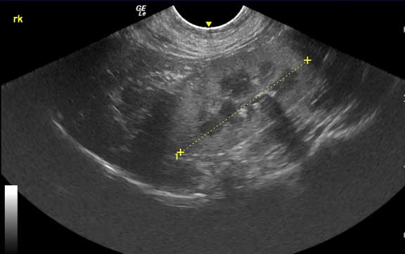
Looks get familiar with the anatomy of the abdomen using ultrasound:
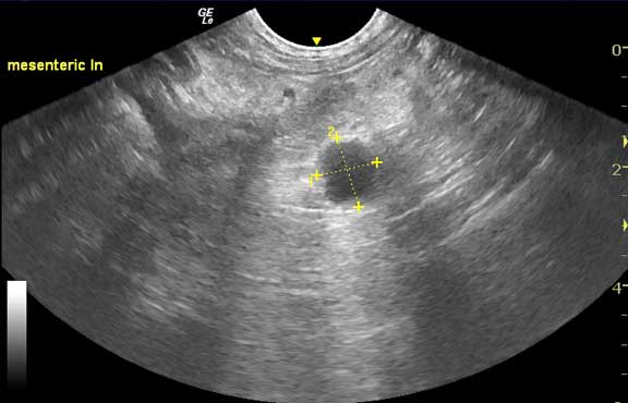
The mesenteric lymph node in the center of the small intestines

The left kidney

The pancreas
The right kidney
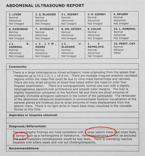
This is typical of a report we get on a dog’s abdomen from our radiologist. It is detailed, and all abdominal organs are checked. Prior to the advent of ultrasound we had to do an expensive and painful exploratory surgery (called a laparotomy or celiotomy) to get similar information. We have the best of both worlds with ultrasound due to less expense and it is non-invasive.

This dog has liver cancer

The liver tumor is demarcated by the yellow arrows
You can learn much more about cancer, including the liver cancer this dog has, at our cancer link. When you go to our individual Diseases Page you will see many more ultrasounds like this.
Ultrasound is highly beneficial in determining if surgery is warranted, and when it is warranted. In the case of the report below, it told us a huge mass in the abdomen seen at X-ray was a spleen that was probably not malignant hemangiosarcoma, that it was probably a benign spleen hematoma, and surgery would probably be curative.
Look at the radiograph of this huge spleen first and you will see why we were concerned about doing the surgery in the first place.
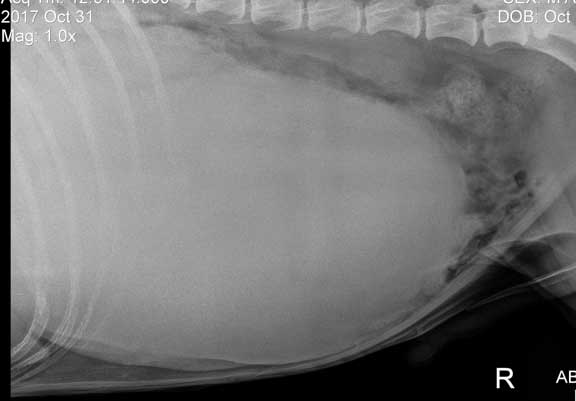
That is 14# of spleen you are gazing at!
The ultrasound gave us a reason to do the surgery, which turned out successful. You can learn all about this case, and the surgery to correct it, at our Spleen Hematoma Page.
At the two week post surgery recheck we have one happy mom (and one 14# lighter dog) glad that we did the ultrasound
Not every patient we ultrasound is sick. This dog is getting an ultrasound in preparation for a C-section.

It is performed in the same way and with the same equipment as a pregnant woman

They look a whole lot different when they come out compared to all that grainy gray stuff on the ultrasound screen while still in the uterus
We do ultrasounds on a wide variety of species like this hamster (we call them hammie’s). With such variability on what is normal or not normal, many ultrasounds of the non dog and cat species need to be interpreted by a specialist.
The lab we use for our blood panels has a division called Antech Imaging Services (AIS). They have a veterinarian named Marie Rush that is board certified in zoo animals to help us in interpreting these more unusual species.

The biggest challenge is getting this wiggly guy with fat cheeks to hold still long enough
Echocardiogram
An ultrasound of the heart is called an echocardiogram. Our radiologist Dr. Ann Reed, the person that performs our abdominal ultrasounds, also does echocardiograms. We more commonly utilize the expertise of Dr. Fred Brewer, our cardiologist, for echocardiograms.

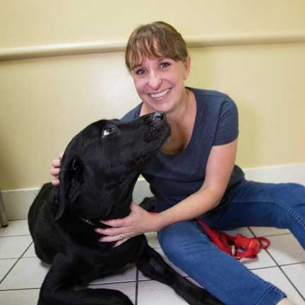
Dr. Brewer teaching one of our student externs

He is teaching her how to identify the valves of the heart

He uses the color doppler to help make a diagnosis
You can hear Dr. Brewer instructing our student in the background as you watch the color doppler reveal the blood flowing through the heart valves
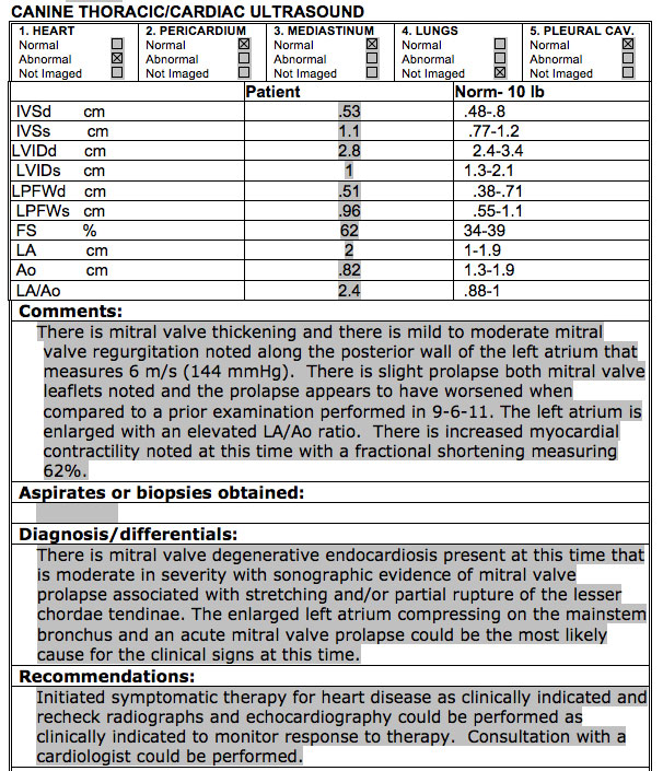
This is the type of report we obtain from our cardiologist Dr. Brewer. After many calculations an echocardiogram gives us more information on the heart than any other modality. It is the gold standard for heart disease in animals.
If you are up to the challenge jump on over to our Heart Page and learn much more about this complicated organ.
Combination abdominal/cardiac ultrasound
On occasion a pet needs an ultrasound of the abdomen and the heart. This is the type of report we receive in this case.
Miscellaneous Ultrasound Procedures
Ultrasound is also used on other organs on occasion. In the veterinary world this usually involves the limbs to look for damage to nerves and blood vessels.