
Introduction
One of the most common surgical procedures we perform is a cat neuter, know medically as an orchectomy. It is performed for several reasons:
- It minimizes roaming
- It minimizes aggressive behavior
- It prevents male cats from impregnating females.
- It minimized urinating in the home (urine spraying).
Male cats are territorial and prone to fighting, which leads to serious diseases, especially viral diseases like FeLV, FIP, and FIV. Neutering minimizes this fighting, helping also to cut down on these serious contagious diseases.
At the Long Beach Animal Hospital we use the carbon dioxide laser for our cat neutering. There is almost no bleeding during the surgery, but most importantly, there is negligible swelling and pain post operatively. Your cat will appreciate this.
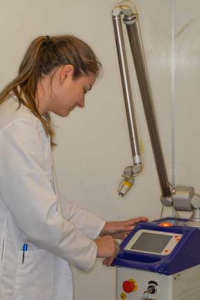
Calibrating the surgical laser just prior to using it
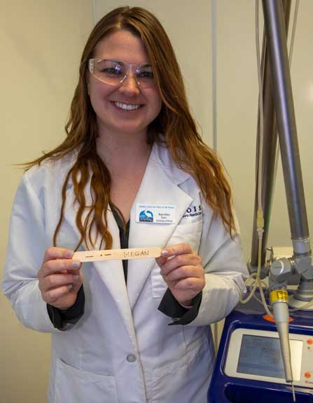
Vet student extern learning how to use the laser by etching her name in a wooden tongue depressor
Sometimes people get a jaded mindset when it comes to routine surgeries like neuters, that are performed by the thousands, especially at low cost spay and neuter clinics. It is a major surgery, and we treat it as such at the Long Beach Animal Hospital, which you will learn about in this page.
Several days prior to any surgery please bring in your pet for a preanesthetic exam and blood panel to confirm your pet is ready for anesthesia. At that time one of our doctors will go over any questions you have.
On the day of surgery we need your cat in the hospital between 7:30 AM and 8 AM. Please take away all food when you go to bed the evening before surgery. Let your pet have water during the night. Do not give your cat anything to eat or drink the morning of surgery.
Our surgeon will call you after the cat neutering surgery is complete and your cat is awake. It can go home in the late afternoon the day of surgery unless instructed otherwise. Please call our office at 4 PM for pickup time, you will be given written post operative instructions then. We are open until midnight if you need to pick up later.
What is cat neutering?
When we neuter a cat we are removing both it its testicles. At the beginning of this page the for reasons for doing this are mentioned. This is different from spaying, which is only done in females, and is the removal of the ovaries and part of the uterus.
Benefits on Neutering Your Male Cat
Minimizing Roaming Behavior
Male cats that are not neutered tend to want to be outside and are territorial and roam. This leads to a significant increase in the chance of it getting a traumatic injury like being hit by a car (HBC) or getting into a fight with another animal (especially another male cat called a Tom), which causes an abscess (called a Cat Fight Abscess- CFA).
It also helps prevent your cat from getting internal parasites (worms) and picking up viral diseases like the Feline Leukemia Virus (FelV) and the Feline Immunodeficiency Virus (FIV).
Minimizing Aggressive Behavior
Male cats are highly territorial and will fight for their territory outside. The cat fight abscess they can get from this fighting can cause high fevers, depression, loss of appetite, and draining abscesses. These cats need sedation, drain tube placement, and antibiotics.

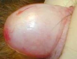
A cat fight abscess over the left eye
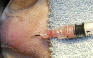
Draining pus from an abscess
Penrose drain tube draining the pus (purulent material) over several days while healing
Preventing Impregnation
A neutered male cat is sterile and cannot impregnate a female, nor does it even have much of an urge to mate.
Reducing Urine Spraying
Since intact (not neutered) male cats are highly territorial, one of the ways they mark their territory is to spray their urine. This is especially true on vertical surfaces in your house. This urine has a very strong odor, and neutering helps minimize this or prevent it completely.
Considerations Before Neutering
Before we perform any surgical procedure it is important that your cat is in good health, has no parasites, and does not have any skin infections. Determining is all part of the pre-anesthetic protocol we follow that you will learn more about on this page, with links to much more detail on all of this.
Where Can You Get Your Cat Neutered?
The Long Beach Animal Hospital has veterinarians that have decades of experience and tens of thousands of surgeries under their belts. In the following surgical section you will learn much more about how we pay attention to detail to minimize the risk of anesthesia and yield a satisfactory outcome.
How is a Male Cat Neutered at the Long Beach Animal Hospital?
Graphic surgical photos later in this page
Anesthesia Process
Pre-anesthetic preparation is important in every surgery we perform, no matter how routine. all of our neuters receive a physical exam prior to surgery.
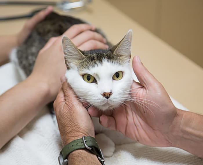
Our pre-anesthetic exam starts at the head and goes to the end of the tail (if they have one)
After this exam will we draw a small amount of blood for an in-hospital pre-anesthetic test. When everything is in order we will give a sedative. This will calm the pet down and make the administration of the actual anesthetic, along with post operative recovery, much smoother. Once a pet is anesthetized, prepared for surgery, and had its monitoring equipment hooked up and reading accurately, the surgery can begin. Cat neuter surgery is a short procedure, and only a small amount of anesthetic is needed.
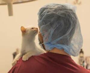
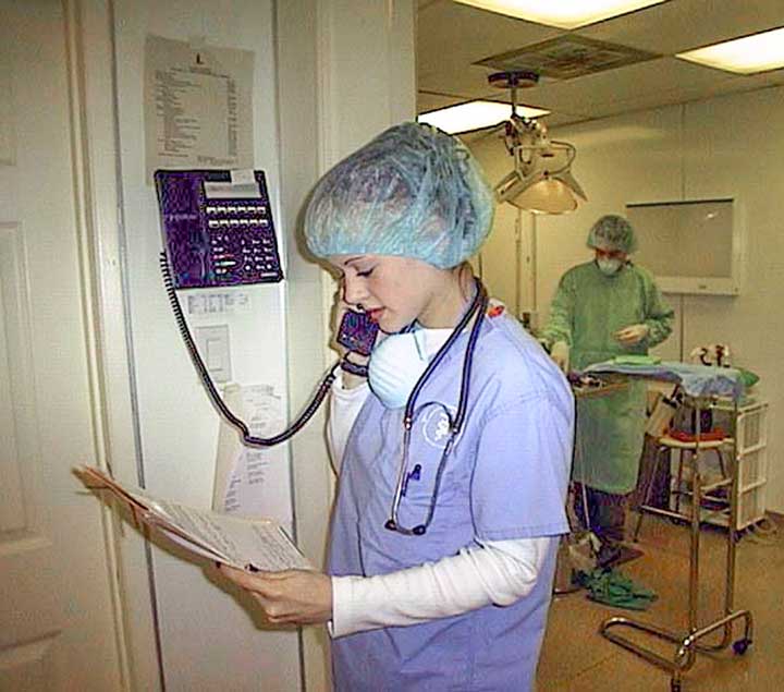
One of our patients being brought into the surgical suite
This is a sterile surgery, and our surgeon starts the pre-surgical process by using special soap to clean his hands

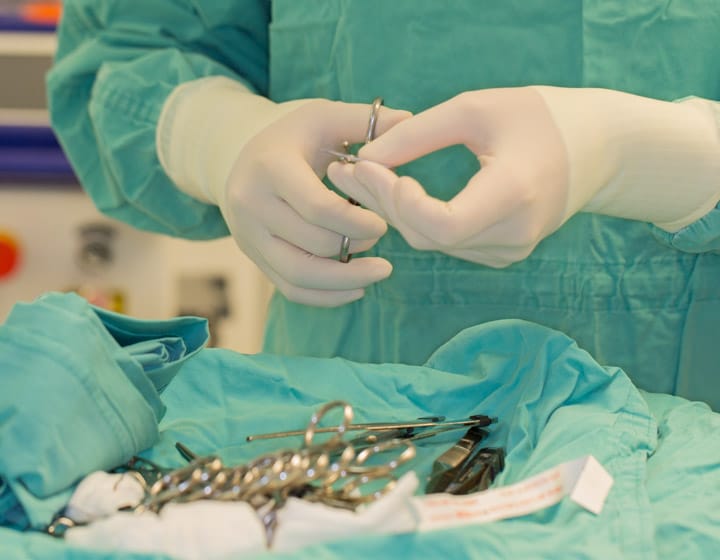
While our patient is being anesthetized our surgeon is already in our surgical suite setting up instruments. Our surgeon is ready to start before our patient is at a proper plane of anesthesia. Once the anesthetist gives the green light the surgery starts immediately. We want our surgeon waiting for his patient, not the other way around. All of this is to minimize anesthetic time.

We keep a close tab on important physiologic parameters for all of our surgeries. Monitors like this give us an early warning of an impending problem.
This machine monitors:
Temperature
Heart Rate
Heart rhythm
Oxygen saturation
Carbon dioxide level
Respiratory rate

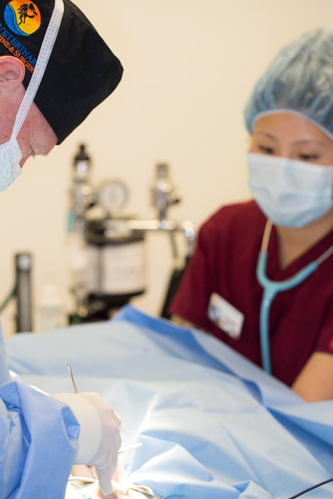
In addition to our monitoring equipment our anesthetist stays “hands on” in monitoring important physiologic parameters
Surgical Procedure
Graphic surgical photos coming up
Case 1: Both testicles are in the scrotum
In general, neutering males cats is a straightforward procedure. Most cats have both testes in the scrotum, making them readily accessible by a scrotal incision. We do not suture the scrotum after the procedure since it heals very rapidly by itself.
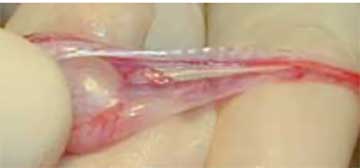
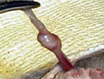
In this picture a small incision has already been made in the scrotum with the laser, and the testicle is visible
Our surgeon has the testicle in his hand which allows visualization of all the internal structures. You can visualize the white and glistening vas deferens at the top of the picture going from the body on the right to the testicle on the left. The vas deferens will be used to tie off the blood supply to the testicle.
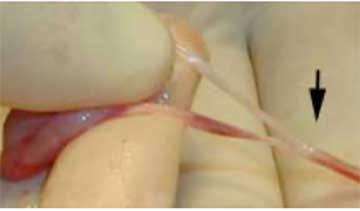
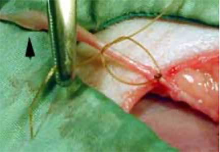
Due to the small size of the blood vessels we can use the natural anatomy of the testicle to prevent bleeding when we remove the testicle. The black arrow points to the knot in the vas deferens made by the surgeon. After several more of these knots are applied the testicle at the far left will be cut off
Cat spermatic cord
Another technique to tie this knot involves the use of a hemostat. This picture shows the beginning of the knot.

After the vas deferens and blood supply are wrapped around the hemostat they are then passed through the center
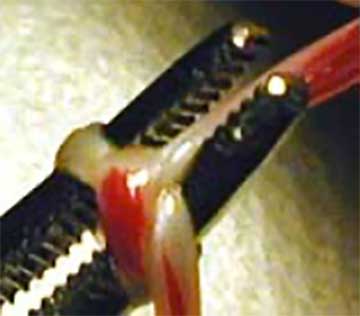
Here is the final appearance after the knot has been completed
At this point anesthesia is stopped and it is moved to recovery to be monitored by our technicians

When fully awake we will call and let you know
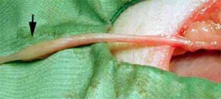
Case 2: One of the testicles is in the abdomen or in the inguinal canal
On occasion both testicles are not in the scrotum. This means that one of them is in the abdomen or in the inguinal canal, which is the passageway through the body wall into the scrotum. Either way, we have to find this testicle and remove it because it will continue to secrete hormones and can potentially become cancerous.
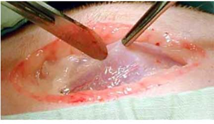
The skin has already been incised and our surgeon is preparing to enter the body cavity. The white glistening structure visible is the section in the center of the abdomen where the abdominal muscles and their tendons meet. It is called the linea alba. We make the incision here due to minimal blood supply and good holding power for sutures
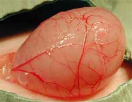
The testicle is not the only thing that resides in the abdomen. This is a picture of the urinary bladder (we hope you didn’t think it was the testicle!) that bulged out of the incision in the abdomen. Obviously, it can interfere with the surgery when it is this full with urine, so we remove the urine before proceeding further.
Testicles that reside in the abdomen are very small because they have atrophied (shrunken in size) due to lack of use. They can be quite difficult to find, and necessitate careful exploration of the abdomen. The black arrow points to the atrophied testicle.
In this case we use suture material to prevent bleeding. The black arrow is still pointing to the testicle to help keep you oriented.
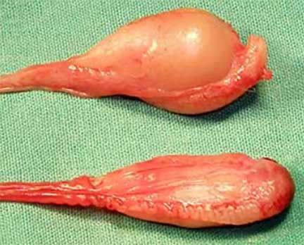
This picture is from another surgery. We have already removed a normal testicle from the scrotum and an abnormal testicle from the abdomen. You can see the difference in size and shape
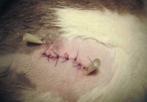
When the surgery is complete we sew up the incision in the muscles and skin, and give a medication for pain. Once the hair grows back it is impossible to tell if surgery was performed.
Benefits of laser surgery over traditional methods
Using the laser has many advantages over using a scalpel blade. These include negligible bleeding during the procedure and post operative pain. We can use the laser to make an incision in the scrotum, which makes the healing process much more comfortable. Our Laser Page has detailed information on the use of the laser for various surgeries.
Post-Surgical Care
Most cats go home the day of surgery quiet but completely awake. We prefer you keep them confined for the night for observation and to give them any pain medication they might need (not needed all the time due to the laser). Keep him away from other pets and children until he is fully recovered.
We have a detailed page on post surgical care of a pet.
What to expect after cat neutering?
Within 1-2 days (oftentimes less due to the experience of our surgeons and the laser) your cat is back to normal eating and activity. If he is not back to normal within 48 hours give our office a call at 562-434-9966.
Some of the following, although rare, might be encountered:
- Grogginess from the pain medication that calms them down
- Lethargy
- Minimal appetite
Check the incision site daily for excess redness or swelling. A small amount is common.
Frequently Asked Questions (FAQ’s)
How long is the recovery time for neutering a cat?
Almost every cat is back to routine activity and appetite within 24-48 hours
Are there any disadvantages of neutering?
The only disadvantage is the risk of anesthesia. With our modern anesthetics and experienced surgeons, this risk is negligible, and the upside to performing the procedure far outweighs the disadvantages.
Do male cats become friendlier once neutered?
Your cats behavior is usually determined as a kitten, and if it is influenced by less male hormones (testosterone) due to neutering, it would be very rare for it to become less friendly, and it will possibly more friendly.
Surgical Services at the Long Beach Animal Hospital
We do a wide variety of surgeries on a vast array of species. Our Surgical Services Page has more information.
We even help with some of the surgeries at the Long Beach Aquarium of the Pacific