
Introduction
Mammary tumors account for 17% of all cat tumors. They are the 3rd most common tumor in cats, after skin and blood cancer. Even though cats get this problem half as often as dogs, almost all of them are malignant. Intact females are at highest risk, although it does occur in males on rare occasion.
Some tumors become so large that they can become ulcerated and painful, leading to the potential of an infection that spreads to the rest of the body (sepsis or septicemia). Why someone would wait for a tumor to get this large before seeking our medical care is beyond us.
It is an accepted fact, proven over many years, that if you spay (ovariohysterectomy) your cat prior to its first heat cycle there is a negligible chance your pet will get this cancer. The longer you wait once your pet starts its heat cycle the greater the chance it will get this problem.”
Even though early spaying in the cat does not seem to yield as much protective effect as in the dog, you can still decrease the incidence of this tumor by up to 60% by spaying early.

Some tumors become so large that they can become ulcerated and painful, leading to the potential of an infection that spreads to the rest of the body (sepsis or septicemia). Why someone would wait for a tumor to get this large before seeking our medical care is beyond us.
Mammary tumors are very common in rats.

Sometimes by the time we see them they are as large as a rat
At the end of this page are two short videos of surgery using the laser to remove a mammary tumor. They are graphic in nature, and not suitable for all viewers.
Anatomy
There are 5 sets (varies from 4-6) of mammary glands in a chain, for a total of 10 mammae. From top to bottom they are called:
- Cranial thoracic
- Caudal thoracic
- Cranial abdominal
- Caudal abdominal
- Inguinal
The inguinal mammary tissue tends to the largest, and produces the most milk. Due to its size this area can look like it has a tumor when in reality it is normal. If you feel an enlargement here one of our doctors should check it to confirm it is nothing more than fat.
The upper mammary glands drain towards the auxillary (arm pit) lymph nodes. The inguinal mammary glands drain towards the inguinal (groin) lymph nodes. The middle mammary tissue (caudal thoracic and cranial abdominal) can drain in either direction. You can learn much more about lymph nodes by following this link.
In the cat there are 4 pair of mammary glands. The cranial gland are the most common ones for tumors to occur.
For a little comparative anatomy fun; manatees, and primates only have two mammary glands.
Physiology
Mammary glands are modified sweat glands. They reside in the subcutaneous (SQ) fat, which is the fat just under the skin but above the muscle. The primary function of the mammary glands is to produce milk and hormones.
Milk contains:
- Water
- Lactose (the milk carbohydrate)
- Fat- much higher in some animals than others, usually in the form of triglycerides.
- Protein- Also varies quite a bit by species. The primary protein in milk is called casein.
- Mineral, vitamins, and enzymes.
Whale and seal milk has 12x as much fat, and 4x as much protein, as cow’s milk. Cow’s milk has less protein and fat than cat milk, which is why orphan kittens and puppies do not do well on it. It takes between 500 and 1000 liters of blood to make 1 liter of milk in the cow.
Numerous hormones are involved in the production of milk:
- Progesterone
- Insulin
- Glucocorticoids (cortisone)
- Prolactin
- Estrogen
In the first week of lactation the milk that is produced is called colostrum. This milk contains antibodies to protect kittens and puppies from routine diseases like Distemper and Parvo.
In the cat there are usually progesterone receptors, the estrogen receptors are not very prevalent.
Classification of Mammary Tumors
Mammary tumors can be malignant or benign. In dogs, up to 50% are malignant. In cats, almost all mammary tumors are malignant (adenocarcinomas). Although there are histologic variations on this, these are the main classifications. The more common ones are at the top of each list:
Benign
- Adenomas
- Mixed tumors
- Fibroadenomas
- Mesenchymal
Malignant
- Tubular adenocarcinomas
- Papillary adenocarcinoma
- Anaplasric carcinoma
- Sarcomas
- Solid carcinomas
- Mixed
Cause of Feline Mammary Cancer
The exact cause of mammary tumors is unknown, although there is a strong correlation to hormones. It has to do with estrogen and progesterone receptors on the tumor. These receptors are present in up to 70% of canine mammary tumors, and 10% of cat tumors. For this reason we tend to stay away from estrogen and progesterone type drugs when treating other diseases.
If your dog is spayed (ovariohysterectomy) before it goes into its first heat cycle, the chances this cat will get breast cancer later in life is virtually nil. A typical female dog will go into heat at 9 months of age, although this varies. If your dog is not spayed until after its first heat cycle the risk of breast cancer can be as high as 8% later in life. Another heat cycle prior to spaying gives a 26% chance of cancer later in life.
Another way to minimize the risk of mammary cancer is to keep your pet at its proper weight.
It is also beneficial to spay a cat early in life. This is especially important in cats because most of their breast tumors are malignant.
Symptoms
The beginning signs of breast cancer can be hard to detect because they are so subtle. Also, mammary tissue tends to hang down hiding any swelling or enlargement.
You should examine your cat weekly while you are playing with it or petting it. Most pets love to have their bellies scratched, which is an ideal time to do your exam. We will show you how to do an In-Home exam next time you come in to check for any problem your pet might have and diagnose and treat it early when there is so much more that can be done to effect a cure.
Run your hands along both chains of mammary tissue from top to bottom feeling for any difference in symmetry. Palpate each gland individually and gently for swelling, discharge, ulceration, hardness, extra warmth, nodules, or discomfort.
Look at each mammary gland, especially each nipple, for any signs of discharge, inflammation, or swelling. Any of the above symptoms are an indication to bring your pet in for us to perform an exam and even run some tests if we think a problem is present. Other symptoms to look for are lameness, swelling of the limbs, or difficulty breathing.
How is a cat’s breast cancer diagnosed?
A thorough approach is needed for a correct diagnosis of mammary tumors. In every disease we encounter we follow the tenet’s of the diagnostic approach to ensure that we make an accurate diagnosis, and also so that we do not overlook some of the other diseases that are common in pets.
Diagnosing some cases of mammary tumors is straightforward, especially if the disease has been present for a significant amount of time before a diagnosis is made. Unfortunately, in these cases the disease can be well entrenched, and malignant tumors have had significant time to spread.
-
Signalment
This tends to be a disease of middle-aged and older unspayed female cats. Even though it can occur it is rare in males.
Some breeds have a higher incidence:- Hunting breeds- retrievers, pointers, and spaniels
- Terriers- Boston, fox, and Airedale
- Dachshunds
- Poodles
- German Shepherds
Some breeds have a low incidence:
- Collies
- Boxers
Siamese cats have twice the risk as other cats, and their tumors tend to me more malignant than other cats. Domestic shorthair cats (DSH) have a higher incidence than other cats also.
-
History
Due to the location of the breast tissue it is easy for an owner to overlook this problem . Pets with early breast cancer do not show the usual symptoms of disease in general. They are usually active, eating well, maintaining normal weight, and have normal bathroom habits. A small tumor that is growing can easily be present for months before an exam is performed. This adds to the problem and can make treatment more complicated.
Some owners find a swelling, discharge, or a growth while bathing or petting their cat. Any suspicious area should be checked by one of our doctors to determine if there is a growth, swelling, or just normal breast tissue.
When the tumor has already spread some pets might have difficulty breathing (dyspnea) due to buildup of the tumor in the lungs, or lameness due to spread of the tumor to the bones. In cats the dyspnea can be due to fluid buildup in the thorax (pleural effusion).


This cat has a mammary tumor at the nipple. This is the only sign of disease it had, and can easily be missed if you are not observant. This problem was easy to spot once we clipped the hair in preparation for surgery.

Other diseases that mimic breast cancer include an infection called mastitis, skin tumors, and an inguinal hernia. In cats the inguinal fat pad can be enlarged and mimic a tumor. Foreign bodies like BB’s (not all that uncommon for a cat to be found with a BB when we take an X-ray) feel like tumor nodules.
Physical Exam
In some cases a swelling or growth is found in the breast tissue during an exam for a different problem, or during a routine wellness exam.
Nodules might be small and solitary, or the whole mammary chain can be affected. Nodules that are adhered to the skin or underlying tissue, are ulcerated, painful, or swollen tend to be malignant. Nodules that are rapidly increasing in size also tend to be malignant. There might be a discharge from the nipple, and your pet might be running a fever.
Here are some typical lesions in a cat


Whenever your pet is placed under anesthesia we perform a thorough exam, including mammary tissue. This is an ideal time because your pet is not moving, it is commonly on its back and we have good access and visualization of the area, and the muscle relaxation allows us to thoroughly palpate small nodules. Your pet can have a malignant tumor and show minimal to no symptoms.

Enlarged lymph nodes due to spread of tumor might also be noted. A lymph node can contain the spread of tumor cells and still appear and feel normal. Cats frequently have the spread of their tumor to the lymph nodes.
One of the typical lymph nodes we will check during an exam are the axillary (arm pit)

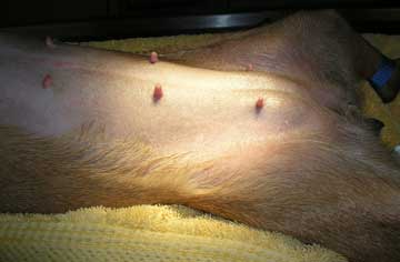
In this picture that cat’s head is to the right, and we are checking the inguinal lymph nodes on the insides of the rear legs. If a malignant tumor has spread through the lymphatic system it can cause swelling of the rear legs due to blockage of the lymphatic drainage system.
Diagnostic Tests
Any pet suspected of having a mammary tumor needs routine tests as the first part of the diagnostic process.
Blood Panel
A CBC (complete blood count) and biochemistry panel should be run on every cat 8 years of age or more, especially if they have any of the symptoms of mammary disease
The CBC checks red and white blood cells. We are looking for signs of infection, cancer, anemia, or excess production of red blood cells. If your pet has mammary cancer it might also have inflammation or a secondary infection. We might get a clue of this from the CBC.

This pet might have an inflammation or infection as evidenced by the increase in the white blood cells. This is called leukocytosis. If the physical exam findings are consistent with an infection then we might put this pet on antibiotics before initiating any other treatment at the moment.
The next part of the blood panel is called the chem or biochemistry panel. It checks the internal organs, along with electrolytes and specific physiologic tests like blood sugar.
Cats with mammary tumors tend to be older, so Geriatric Diseases are more prevalent. Since surgery is usually a major part of treatment we need to make sure the internal organs are ready for anesthesia. This is particularly true of kidney and liver disease. Some pets with mammary cancer will have a high calcium level as seen on this panel.

This pet has some abnormalities, especially the low protein level, that need to be addressed prior to surgery
In some cases we might run a clotting panel looking for any signs of a disease called disseminated intravascular coagulation (DIC). This can occur when there is an inflammatory carcinoma.
Urinalysis

The urinalysis on this pet is normal
Cytology
In this test we insert a tiny needle with attached syringe in the mammary tissue. It is a tiny pin prick, and is the same kind of needle we use to give vaccines. Some cells are aspirated into the syringe and then gently pressed on to a microscope slide.

In some cases we use this test, especially if it is difficult to differentiate inflammation from an actual tumor. Its also gives us an idea of just how malignant or non-malignant the tumor is, so we can adjust our surgery accordingly. In cats we assume the tumor is malignant and usually skip this test and go right into surgery.
Even though it can be a useful test, it only looks at a small portion of the mammary tissue. So it is used only as ancillary information prior to surgery and not to make a final diagnosis. Also, multiple tumor types might be present, and you can make the wrong interpretation with just this test.
Aspiration of a local lymph node can also be helpful to detect evidence of any spread of a tumor. In cases of extensive mammary involvement, usually the whole chain, we might completely remove the lymph node that drains that area. This gives the pathologist much more tissue to work with to ascertain if there has been a spread of the tumor.
In cats that have fluid buildup in their thorax we can submit this fluid for cytology also.
Radiography
Radiography (X-Rays) are a very important test prior to surgery because up to 50% of the dogs with malignant breast cancer have spread of the disease to the lungs at the time of their exam. We need to confirm that there is no spread (metastasis) of the tumor to the lungs or else surgery might not be indicated. We take 3 different views of the chest to determine if the lungs are clean.

In this chest radiograph we have placed black arrows at some of the white and round areas that are the spread of the tumor in the lungs. Compare it to the normal radiograph below if you need to.

Normal chest radiograph

This is what a mammary tumor under the skin looks like on a radiograph. It is that round white object on the bottom
Some cats will show signs of difficulty breathing. It can be subtle, so it behooves you to spend some time every day observing your pet for any changes that indicate a problem. A normal respiratory rate in a cat at rest is 30 times per minute. Check your cat as part of your routine In-Home exam to see what is normal for your pet. What you are looking for is a change in the respiratory rate, and if it increases, we should examine it.

This is a normal cat chest radiograph. Notice the large and normal black lung area.

In this radiograph from a problem cat there is fluid throughout the thorax and you cannot see normal black lungs. The lungs have collapsed due to the fluid in the thorax. The only lung tissue you see is the slightly dark leaf-shaped structure towards the top of the thorax.

After oxygen therapy for stabilization we drained some fluid off the thorax. This caused an immediate improvement in breathing. A radiograph taken soon afterwards shows improvement as evidenced by the increase in the normal amount of black lungs visualized. When this fluid appears due to the spread of a malignant tumor from a mammary gland the prognosis is poor.
In some cases a radiograph of the bones will show spread of cancer. If a radiograph is taken of the abdomen some malignant cancers will show an enlarged sublumbar lymph node. Ultrasound is beneficial here in assessing local lymph nodes and abdominal spread from a malignant mammary tumor.

This abdominal radiograph shows the location of where the sublumbar lymph node is normally located. It is not apparent in this view, so it is not enlarged. The K stands for kidney and the B stands for urinary bladder. Ultrasound tends to be a more accurate way to assess abdominal lymph node enlargement when compared to radiography.

Can you see the enlarged sub lumbar lymph nodes in this radiograph?
Response to Therapy
One of the tenets of the diagnostic process is whether or not a treatment that is instituted actually corrects the problem. Surgery is the main form of treatment, so response to treatment does not apply as much as to other diseases that are more medical in nature and treated with drugs.
Treatment
The treatment of choice for mammary tumors is surgery. Chemotherapy, radiation therapy, nor hormonal therapy have any proven benefit.
If the gland is infected we might use antibiotics to reduce the swelling and inflammation. This will allow us to see the margins of the tumor more readily during surgery.
When your pet is relaxed under anesthesia, and the hair is clipped away prior to surgery, we will examine the mammary glands again. It is not uncommon to discover a small tumor that was missed during the routine exam.
Any pet under anesthesia is closely monitored with the latest equipment
Our anesthesia page (link above) has detailed information on how we anesthetize the wide variety of animals we care for at Long Beach Animal Hospital.
Once our diagnosis and ancillary tests are complete we will remove the mass surgically. Depending on the location, size, duration, species, and physiologic status of your pet, we might do a lumpectomy or remove part or all of the chain. In extensive cases we might have to remove one chain of tissue in a first procedure, then the other chain several weeks later when the first chain has healed. Since cats usually get malignant tumors it is common to remove the whole chain on the affected side.
We routinely use our laser for this surgery. This dramatically minimizes post operative bruising, discomfort and swelling. We can’t emphasize enough how important the use of the laser is in this surgery.
Prior to our laser these cats would have extensive bruising and swelling of the sensitive mammary tissue. We would place many sutures under the skin to prevent fluid buildup and discomfort.
We no longer need to with the laser. Cats that have this surgery, even when a radical surgery is performed, routinely go home the same day and have minimal discomfort. When laser is used with routine pain medication your pet will usually be eating and resume normal activity within 12-24 hours.

We use the laser to remove these tumors due to its tremendous ability to control bleeding during surgery, and swelling and pain after surgery. As you can see from the picture above there is no blood in this dissection of this mammary cancer. Without the laser it would have been very bloody.
In all these surgeries we remove a wide margin of tissue to ensure we removed all of the tumor. In all cases our goal is to remove all of the tumor and get what are called “clean edges” by the pathologist. This means there is no microscopic signs of tumor cells in the tissue submitted for analysis. This makes for a much better prognosis.

This is a typical mammary tumor in a cat prepped, draped, and ready for surgery

A wide surgical incision is made with the laser to makes sure we have removed all of the cancer in this cat. The lack of bleeding due to the laser is obvious.

What the suture site looks like when we are finished. The rubber tube is called a penrose drain tube.
It is used to decrease swelling during healing, and is removed in 3-5 days. In most of our laser surgeries we do not need to put this tube in since there is no post-operative swelling.

Before our patients wake up we use the Cold laser to minimize swelling at the incisions site and aid the healing process
We use it in many of our surgeries where an incision is made in the skin. Here it is being used after a dog neuter.

We make sure our patients are comfortable after a mammary chain removal. This involves pain medication prior to, during, and after surgery. It also involves the use of the laser during the surgery and local anesthetic at the incisions site. Finally, after we use the Cold Laser on the incision site, a comfortable wrap is put around the chest with padding underneath.
Since cats frequently get malignant tumors we commonly remove the whole chain of mammary tissue on the affected side. We might also remove the closest lymph node to look for metastasis.

This is a report on a cat with a malignant breast cancer
When we remove the mammary tissue on one whole chain there is a long incision. With the use of the laser and routine surgical and post surgical pain medication these cats recover rapidly from surgery.

Click on the link below to see a laser lumpectomy surgery on a cat. Notice how little bleeding there is when the laser is used.
This next cat has more gland involvement and requires more surgery. Notice how diseased the tissue appears and the lack of bleeding when using the laser. As the surgery progresses you can see milk coming from the gland.
Click here to learn more about the laser and how it is used in many types of surgeries at our hospital.
Post surgery treatment
Diseased mammary tissue that is removed during surgery is submitted for histopathic analysis. The pathologist will determine the type of tumor and will also stage it. Stages usually go from 0 – 3.
- Stage 0- Tumor cells are limited to the ducts within the mammary tissue
- Stage I- Tumor cells are in the ducts and the supportive or framework tissue of the mammae (called stroma)
- Stage II- Tumor cells are in the blood vessels, lymphatic tissue, or regional lymph node
- Stage III- Tumor cells have spread through the body- usually lungs or bones
Chemotherapy is used when we cannot remove all of the tumor of if your pet has inflammatory carcinoma. Chemotherapy for feline mammary cancer tends to be unrewarding. Some of the drugs we use, which should be under the direction of a veterinary oncologist, include:
- Doxorubicin
- Carboplatin
- Mitoxantrone
- Cyclophosphamide
In some cats the tumor is not resectable. This occurs in inflammatory carcinomas. In these cases we used what is called palliative therapy. We attempt to keep them comfortable with antibiotics, pain medication, fluids, assist feeding, good nutrition, and lots of TLC.
Prognosis
Cats with malignant tumors usually do not survive more than a year. Their tumors grow rapidly and spread to the lungs early, usually before a pet owner is aware and brings them in for diagnosis and treatment. Prognosis for a cat depends on 5 factors:
Tumor size
If there is spread to the lymph nodes
Histologic grade:
Stage I- > 24 months
Stage II- 12-24 months
Stage III- 4-12 months
Stave IV- 1 month
Invasion of the lymphatics that drain the gland
Siamese cats have a poorer prognosis than domestic cats

This is the report on the cat with the nipple that was inflamed. We showed you the picture of this tumor earlier when we talked about how easy it is to miss some of these tumors.
Return to Feline Diseases Page.