
An important diagnostic modality we use to make an accurate diagnosis on a bird with a fractured (broken) leg is radiography, more commonly known as x-rays. Just like the many other Diagnostic tests we perform, x-rays are important in veterinary medicine since our patients do not routinely tell us where they are having a medical problem.
You can learn about the basics of fracture repair in birds from the Orthopedic Surgical Techniques chapter in Avian Medicine.
Due to the fact that birds have small, and sometimes hollow bones, it is not unusual for them to break them due to trauma. Falling off a perch, being injured by another animal, or even being stepped on by its owner are some of the more common causes of a fracture.
Sometimes there are underlying problems causing the bones to be weak and susceptible to fracture during normal activity. The tibiotarsal ( tibiotarsus or shin bone) is the one most commonly fractured.
Bird leg fractures heal quickly, often faster than in humans and other animals. A stiff splint is usually used to keep the bone still so it can heal properly. For more complicated fractures, surgery may be needed to put in supports above the “splinting” point.
This page shows a tape splint on a simple fracture. This splint is light and stable, perfect for a small bird like a budgie (parakeet) or a cockatiel. At the end of this page you will see fractures that need surgery, with a link to seeing the full surgical repair of a fractured femur (thigh bone) in a rabbit at our hospital.
At the end of the page are radiographs of birds that need more than a tape splint. There will be links to show how corrected the problem.
A fractured leg (broken bone) in a bird can be an emergency. They have very little blood, and blood loss can occur from the bone. They are also susceptible to pain and shock, which can be life threatening.
The Long Beach Animal Hospital, staffed with emergency avian vets, is available until the evenings 7 days per week to help if your pet is having any problem, especially if you suspect a broken bone, it is breathing hard or bleeding. Think of us as your Long Beach Animal Emergency Center to help when you need us for everything from minor problems to major a major emergency.
We serve all of Los Angeles and Orange county with our Animal Emergency Center Long Beach, and are easily accessible to most everyone in southern California via Pacific Coast Hwy or the 405 freeway.
If you have an emergency that can be taken care of by us at the Animal Emergency Hospital Long Beach always call us first (562-434-9966) before coming in so that our veterinarians can advise you on what to do at home and so that our staff and doctor can prepare for your arrival. To learn more please read our Emergency Services page.
We work closely with our radiologist Dr. Ann Reed, along with imaging specialists from Antech Imaging Services (AIS) to aid us in using X-rays for diagnostic purposes on our avian (bird) patients.
After you learn how we fixed this bird’s broken leg come back here and learn much more about how we take radiographs (x-rays) on a wide variety of animals from our How To Read a Radiograph Page.
Diagnosis
Most birds that have broken legs will not bear weight on the affected leg. Most fractures can be palpated by one of our doctors, although a bone can be fractured without any obvious evidence during examination. Taking a radiograph is one of the best methods to determine if a fracture is present. Look at the following one and see if you can find the problem.
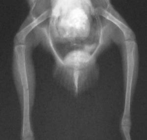
Do you see the fracture in this view?
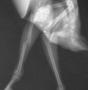
The fracture is more apparent in this side view. This emphasizes the importance of taking two views. This fracture is classified as a mid shaft transverse fracture of the tibiotarsal bone.
Splinting
Most tibiotarsal bones heal well when splinted with special tape. We usually keep the splint on for one month, although this varies. On occasion we need to perform surgery for proper stabilization. This is more common when there is a fracture of the femur (thigh bone).
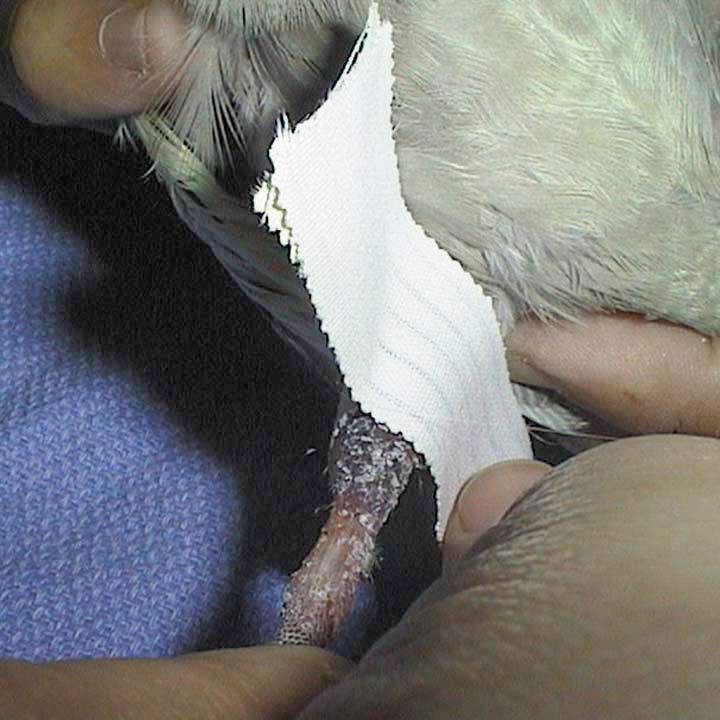
The first layer of tape is applied directly to the skin. We use a type of tape that will cause minimal irritation to the sensitive skin during the healing process and when we remove it after the bone has healed.
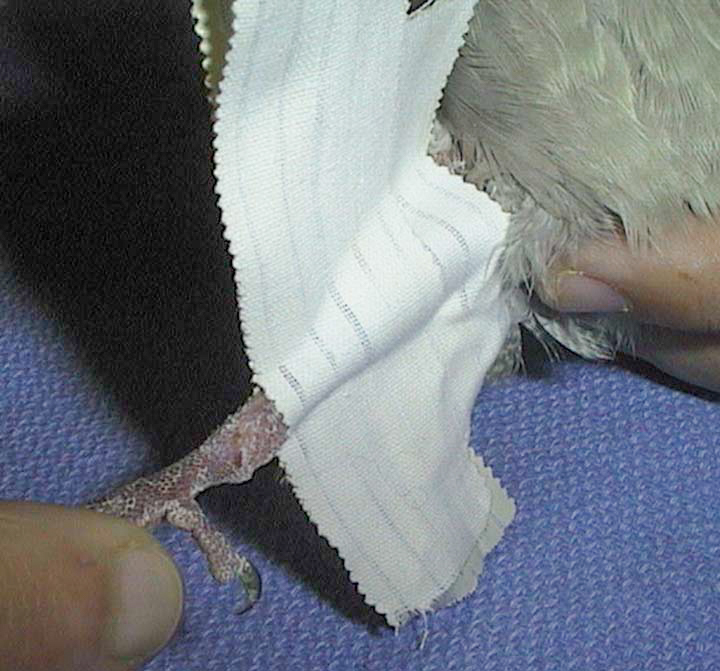
These tape strips are molded over the leg to provide the beginning stages of stability. Several pieces are used to cover above and below the fracture.
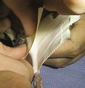
The next layer of tape is waterproof and much stronger. Several strips are used to provide the necessary stability.
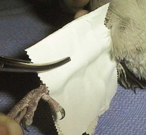
When all the layers of tape are applied a hemostat is used to gently mold all the layers tight up against the bone. Now the fracture site is stable and the bone can begin the healing process.

One our doctor feels the bones are lined up properly and the fracture site is stable, the excess tape is trimmed
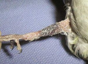
The splint is trimmed for easier mobility. With the fracture stable this bird will feel substantially better, and start bearing weight very soon. It needs to remain quiet and not climb or play excessively during the healing process. The foot should be checked daily for swelling and the splint should be kept clean and dry (of course it is OK to sign the splint). Weekly rechecks by one of our doctors will ensure the splint is secure and the foot is not swelling.
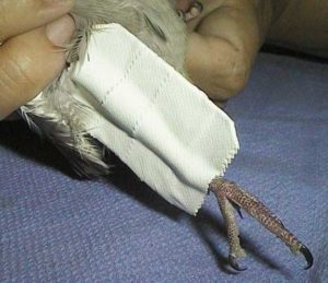
The final appearance of the light but stable tape splint
This budgie also had a broken tibiotarsal bone that was repaired with a tape splint. His bones are smaller than the cockatiel above so gentle handling is a must.
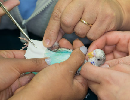
Even though he is small it is a several person job to put the splint on him

He stood on the fracture leg immediately after the splint was applied

He was grateful we took away his pain and started playing with us right away
For comparison purposes this is a cast put on a wiggly 6 year old boy named Mike. Your bird needs the same kind of attention during its healing process. Make sure it gets plenty of rest and good nutrition, and remove perches initially so it can not climb around the cage. Putting something soft in the bottom of the cage is also needed.
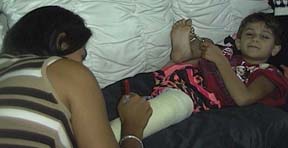
As you can see, one of his fans is signing the cast while he enjoys some R & R
Surgical Fractures
Some tibiotarsal fractures will not heal with a tape splint. The following two radiographs are typical of bird fractures that require surgery, one with a fracture tibiotarsus, the other with a fractured humerus. They are both from our Wildlife Program, and you can see both cases in more detail on our Wildlife Care page.

An albatross from our Wildlife Program
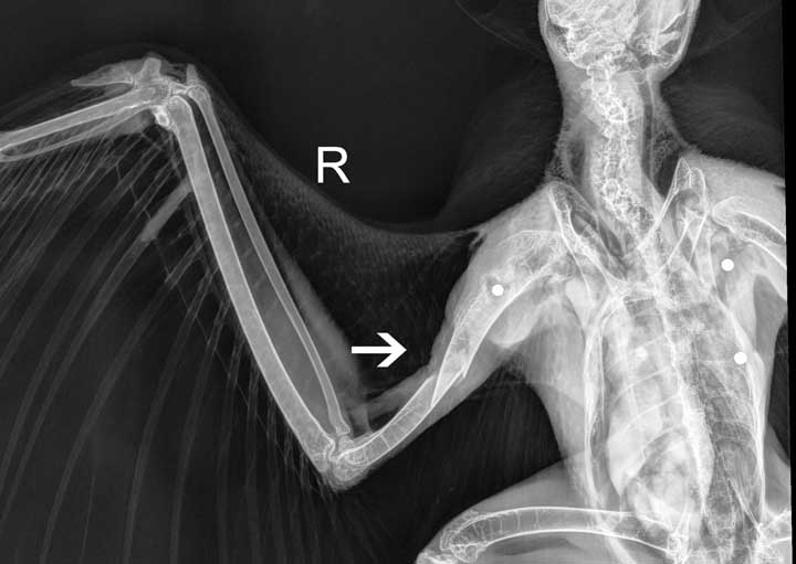
A red-tailed hawk with a fractured humerus. It was successfully released in northern California.
We would like to use plates on birds fractures like other animals. Bird bones are too thin, and the plate is too heavy, to use in smaller birds if they are to fly again. We would need to go back in surgically and remove the plate, which is an added risk.

This is how we put one of those plates on a pomeranian with a fractured radius. It was never removed, and healed 100%.
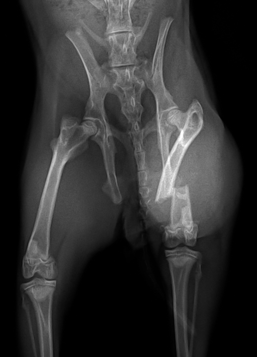
Rabbit with a transverse mid-shaft fracture of the femur (thigh)
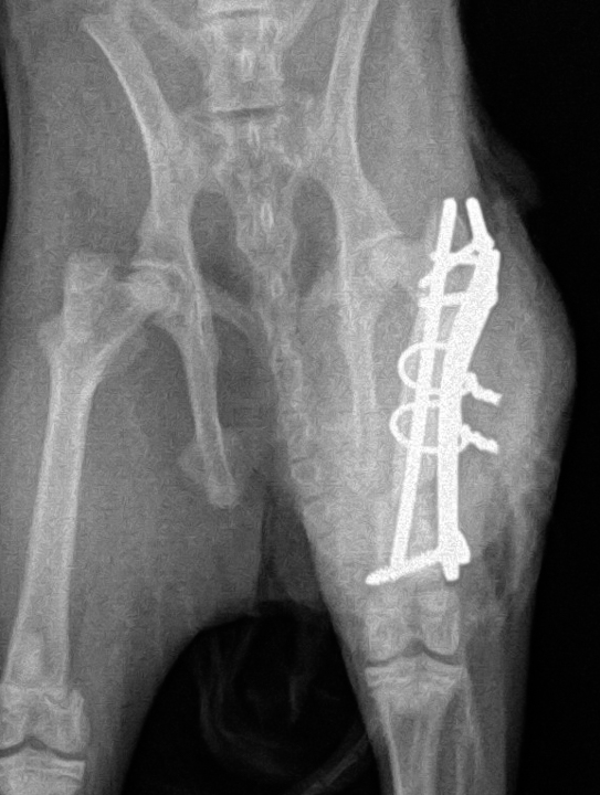
What the plate, screws, and circle wires look like radiographically after the surgery. Click here if you want to see the full surgical repair of this rabbit fracture.
Return to our Avian Diseases Section