
Introduction
Pets that roam outdoors are prone to numerous traumatic injuries. Fighting with other animals, and getting hit by a car, are some of the more common ones. This page shows a surgical procedure to correct an abdominal hernia in a cat named Sundance that was hit by a car.
What is a Hernia?
In this case the hernia was caused by a blow to the abdomen by the car, causing an abdominal hernia. The blow was strong enough to tear a large hole in the abdominal muscles that surround the abdomen. The small intestine went through this hole and was trapped between the muscle and the underside of the skin. This needs to be corrected because the blood supply to the intestines can be compromised while entrapped in this abnormal location. This will cause a segment of the intestines to die with subsequent loss of life as a possible outcome.
Symptoms of Hernia in Cats
There are many different kinds of hernias with variations in the symptoms bases on the type of hernia and duration. This page goes into detail on an abdominal hernia caused by trauma. Symptoms for this type of hernia tend to be:
- Bruising at the affected area
- Lethargy
- Pain
- Reluctance to be petted
- Staying in one place
- Vocalizing
- Poor appetite
- Resistance to being petted
Different types of hernias in cats
In addition to the abdominal hernia in this page, there are other types of hernia’s in cats:
Umbilical
This is a minor hernia that occurs at the umbilicus “belly button”. Usually there is only a small amount of fat that has protruded through a minor hole in the muscles of the abdomen. This problem is easily corrected, and that is usually done during a spay of a female cat or a neuter of a male cat.
Inguinal
This hernia is also rare in cats, and occurs when abdominal contents go into the inguinal canal in the groin area.
Hiatal
This is rare in cats
Diaphragmatic
In this hernia abdominal organs like the liver have torn through the diaphragm and are now in the chest cavity. This is usually caused by trauma like being hit by a car or similar trauma.
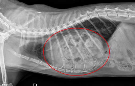
Under the red circle is the heart that is surrounded by by intestines in this diaphragmatic hernia. You can see some feces in the large intestine in the lower right. These should be much further back, and have moved there because they were pulled there from the other intestines that are now in the chest (thorax).
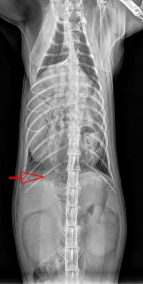
In this VD (venture-dorsal) view the red arrow points to those same large intestines
How is a Hernia in Cats Diagnosed
Diagnosis with Radiography
Our physical exam gave us an indication there was a hernia when we palpated the abdomen. To help us confirm the diagnosis we took a radiograph. Radiographs are taken for numerous reasons on every pet that is hit by a car. These pets can have trauma to the chest, broken ribs, herniation of abdominal contents into the chest, and ruptured internal organs like the urinary bladder. The radiograph helps us determine if any of these problems exist. In this case, the radiograph helped verify that Sundance had an abdominal hernia. He also had a fractured pelvis which would heal on its own if he was confined and rested for one month.
In addition to an abdominal hernia, which you will learn more about soon, animals that are hit by cars also can get a diaphragmatic hernia. In this hernia, the abdominal organs like liver or stomach have literally torn through the diaphragm (muscle of respiration) and are sitting in the chest. Needless to say, these are serious injuries. We will show you pictures of this at the end of this page. Lets look at how we make a diagnosis of a diaphragmatic hernia with radiographs.

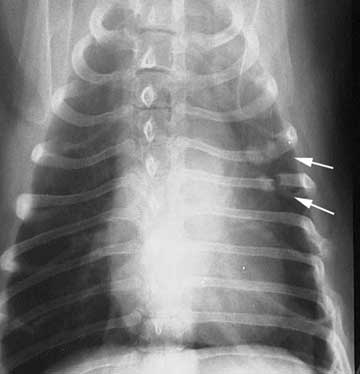
The arrows are pointing to a normal diaphragm. The lungs are the dark areas. Use them for comparison to the abnormal radiograph that follows.

This is the radiograph appearance of a cat with a diaphragmatic hernia. Several abnormalities are apparent:
The windpipe is pushed upwards to the top of the chest
The lungs are not black throughout the whole chest
The diaphgram is not visualized at the bottom
The heart seems huge because the liver and stomach are pushing up against it from below

A different case with a diaphragmatic hernia. It just looks like a big and round heart, but it has a diaphragmatic hernia like above.

This dog has 2 fractured ribs at the arrows. Can you see them?
This view of the same chest might help a little. The fractures could easily be missed in the above view, verifying how important it is to take 2 views.

This is a radiograph of cat with a fractured pelvis that is more severe than the one Sundance has. Do you see the fracture on both sides?
Presentation
Sundance was presented to us with a history of being gone for 5 days and lethargy. Most cats that are hit by a car are in a state of shock and can die if not treated with intravenous fluids. Sundance is lucky he survived being hit by a car without any shock therapy.
Our exam revealed a swollen and bruised area just under the skin in the right inguinal area, which made us suspicious of a hernia. Bruising is very common in such small animals that have been hit by a car, so it does not necessarily mean there is an abdominal hernia.
Treatment of Cat Abdominal Hernia
Anesthesia
Any pet that has been traumatized so severely that it has a hernia has an added anesthetic risk. We take special precautions to minimize this risk.

We keep a close tab on important physiologic parameters for all of our surgeries. Monitors like this give us an early warning of an impending problem.

Once our surgeon has scrubbed up and is in a sterile gown, gloves, and mask, the surgery begins
Surgery Process
In most hernias we make an incision directly over the hernia and proceed to make the repair. In this case, the hernia area had extensive swelling due to the fact that it had been present for several days before we saw Sundance.
Also, the herniated area was near the area where we routinely make an incision to enter the abdomen for an ovariohysterectomy (spay). In this case it was decided to make an incision directly in the center of the body like a spay surgery, and repair the hernia through this incision.
Lets get our orientation before we show surgery pictures. Our patient is on her back, with her head towards the top, and the abdomen clipped of hair. Our patient is already under anesthesia.

The white arrow shows the area of the hernia on the inside of the right rear leg. This is called the inguinal area. It is difficult to visualize the swelling from this view.

This second arrow in the middle of the body shows the location of our incision into the abdomen

The incision directly in the middle of the body was much longer than our typical spay incision. You can see our surgeon starting the incision.
We dissect through the tissue under the skin (called the subcutaneous tissue) until we encounter the rectus sheath, an area where the abdominal muscles come together. This area is very tough, and is used to hold the abdominal muscles together when we suture our patient back together.

The rectus sheath can be seen here as the large white glistening area between Dr. P’s finger. A horizontal incision is made directly through this layer in order to enter the abdomen and find the hernia.

The tear in the abdominal muscles was 4 inches long. It can be visualized here as the horizontal opening towards the bottom of this picture, just under Dr. P’s finger. A large segment of the small intestines was found caught in this hole, and was gently removed just prior to this picture.
Intestines do not belong in this area, and are easily damaged when trapped in an opening this size, especially for 5 days in this case. In this picture Dr. P is carefully checking them to make sure their blood supply in intact.

Here is a different case showing the bruising and compromised blood supply that can occur when the intestines are trapped

Intestines are not the only organ that can be bruised during abdominal trauma or a hernia. The kidney on the left has been traumatized as evidenced by the severe bruising when compare to the other kidney.
A special suture is used to sew the herniated muscle opening shut. It will provide the strength needed to hold the muscle together until healing is complete. Eventually the suture will dissolve.

You can view the partial closure of the opening in this picture as Dr. P sutures the muscle from right to left in the picture.

The muscle closure is now complete. The hernia was so large that additional sutures were placed over this layer for added strength.
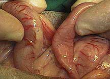
The intestines were not the only abdominal organ trapped in the hole in the muscle. The omentum is tissue that naturally resides in the abdomen. When an abdominal organ is traumatized, as the intestines were in Sundance’s case, the omentum migrates to this area and covers the injured tissue to help in the repair process.

In this picture our surgeon is trimming off a piece of omental tissue that is discolored at the tip
It is important to check all the abdominal organs for injury. After the unhealthy omental tissue seen above was trimmed, Dr. P methodically went through all the abdominal organs and checked for injury.

Here he has exposed the urinary bladder and is looking for any signs of problem
Sundance had no other abdominal organ trauma, so his rectus sheath and skin were sutured back together.

Here is the 7 inch incision in his abdomen after it has been sutured closed. These sutures will be removed in 7-10 days.
At this point in the procedure Sundance was given a pain injection and monitored carefully by our nursing team. He went home the next day and eventually made a full recovery. He was lucky this time, and certainly used up more than one of his nine lives!
Necropsy Photos
To give you a better understanding of anatomy we have some photos taken at necropsy of a diaphragmatic hernia on a pet that did not make it.

This thin, fan shaped, and very strong muscle is the diaphragm. The front side of this muscle is facing the abdomen. On the right side of this muscle is the thorax (chest). You can see the reddish colored liver at the bottom of the picture.

This one has been torn at the bottom

This liver has gone through a torn diaphragm and is now in the thorax. The vertical white line on the left is where the diaphragm was. To the left of this white line is the abdomen, where the liver normal resides. To the right of this line is the thorax, which now contains the liver, when it should not.

When you pull the liver away you can see the heart and lungs
When abdominal organs are in the thorax they take up space and prevent the lungs from expanding. The lungs might also be bruised, called pulmonary contusion.

The darker areas of these lung lobes have pulmonary contusion