
Disk disease occurs in cats and dogs, with the occasion problem in other species. This dog pinched nerve problem is similar to what happens in us people.
Since it occurs mostly in dogs, this page will emphasize disk disease in dogs, although the diagnosis and treatment of disk disk applies to cats and other species as well. A pinched nerve in dogs is similar to a pinched nerve in cats and other species.
Even though it occurs more often in dogs, cats can get it also. This cat is getting laser therapy on its spine, you will learn more about this later in our page.
Breeders have selectively bred dogs over many years to obtain a certain look or to enhance specific behavioral attributes. One of the more significant tradeoffs with this type of breeding is the propensity for some breeds to have problems with their bone structure. In some dogs this affects the spinal cord.
At a minimum it causes discomfort, but unfortunately, it can cause more severe problems, including complete paralysis. When it occurs it is called intervertebral disk disease (IVD). It is one of the most common diseases causing paresis and paralysis of the rear legs in dogs. It is estimated that up to 18% of dachshunds will have this problem. IVD occurs in cats, but not as commonly as dogs.
In though it is not a common, a pet with IVD can be in severe pain and go into shock. Shock is a medical emergency requiring immediate veterinary care. The Long Beach Animal Hospital, staffed with emergency vets, is available until the evenings 7 days per week to help if your pet is having any problems, especially shock, pain, breathing hard, or bleeding.
Think of us as your Long Beach Animal Emergency Center to help when you need us for everything from minor problems to major a major emergency. We serve all of Los Angeles and Orange county with our Animal Emergency Center Long Beach, and are easily accessible to most everyone in southern California via Pacific Coast Hwy or the 405 freeway.
If you have an emergency that can be taken care of by us at the Animal Emergency Hospital Long Beach always call us first (562-434-9966) before coming. This way our veterinarians can advise you on what to do at home and so that our staff and doctor can prepare for your arrival. To learn more please read our Emergency Services page.
One of the most significant treatment modalities for IVD is called VNA, especially since no drugs are involved. We will talk about it more in the treatment section.
Neuroanatomy and physiology is a world unto itself, and is far too complex to cover completely in this page. It will be summarized and generalized for much easier understanding. It is helpful to be aware of some of the medical terms involved with IVD:
| analgesia- lack of feeling or pain | hemi paresis or paralysis- problem with the front and back limb on the same side. |
| ataxia-incoordination | para paresis or paralysis- problem with both pelvic limbs |
| paresis- partial paralysis | mono paresis or paralysis- problem with just one limb |
| plegia- complete paralysis | tetra paresis or paralysis- problem with all four limbs |
Vertebral Anatomy
The dog has 31 vertebrae:
- Cervical (neck)- 7
- Thoracic (chest) – 13
- Lumbar (lower back) – 7
- Sacral (pelvis) – 3 (fused)
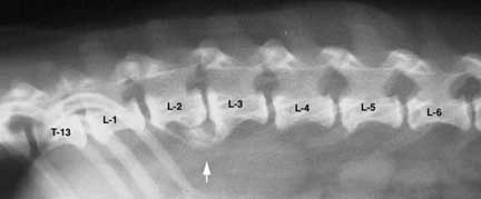
Let’s go on a tour of this anatomy by looking at overlapping radiographs:
The 7 cervical vertebrae are quite flexible, for obvious reasons. Disk disease can occur anywhere in the cervical vertebrae. C-1 and C-2 are called the atlas and the axis. There can be an instability in this area in large dogs that will cause neurologic problems. The term breeders use for this is “wobblers”. This is a pinched nerve in dogs neck.
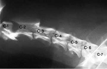
C1 and C2 are the atlas and axis vertebrae
As the cervical vertebrae become the thoracic vertebrae they go past the shoulder (S). The nerves that come off this cervical-thoracic junction at the shoulder are called the brachial plexus (you cannot see nerves on a plain radiograph). They innervate the front legs on each side.
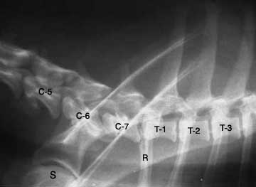
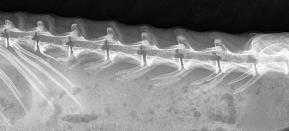
Each of the thoracic vertebrae corresponds to a rib (R) on each side of the chest
As we continue down the thoracic vertebrae you can visualize how high their dorsal spinal processes are. Also notice how these processes start to get smaller as we get closer to the lumbar vertebrae.
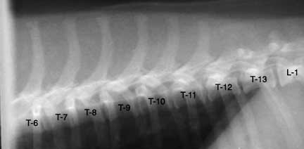
Thoracic vertebrae in this area do not typically cause disk disease
Moving towards the end of the thoracic vertebrae we come to what is termed the thoracolumbar (T-L) junction. As we pass into the lumbar vertebrae we have now made our way into the lower back.
It is a very common area to have disk disease
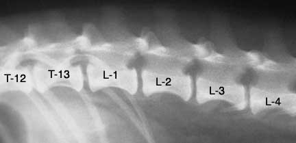
The 7 lumbar vertebrae eventually lead into the sacral vertebrae. The fused sacral vertebrae are hard to visualize because they are within the pelvis. After the sacrum we are at the tail. The section between the last lumbar and first sacral vertebrae is called the lumbosacral (L-S) junction.
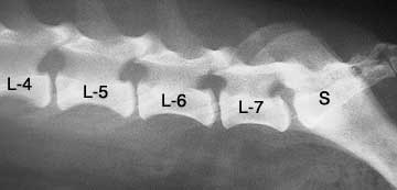
Disk disease and arthritis at the L-7 and S-1 junction can be quite painful
For a fun comparison, let’s look at the lumbar radiographs of other animals besides dogs and cats. See if you can guess before looking at the answer below the radiograph picture.
This is a rabbit lumbar spine. Notice any differences?
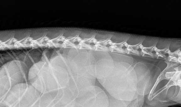
This is from an iguana. You did see the eggs, didn’t you? Click here to see the surgery on how we removed them.
Spinal anatomy
The spinal cord is an extremely sensitive part of the nervous system. In essence, it is an extension of the brain. As the spinal cord moves from the brain down to the tail it sends out nerve branches (called nerve roots) that go to various organs.
Some of these nerve branches bring sensation back to the brain. If you are painfully pinched on your skin it is the nerve branches in your skin that relay a feeling of pain from the skin, to a local nerve branch, and eventually along your spinal cord to your brain.
At the same time that these nerve impulses are going to the brain to tell you it hurts, there are other nerves in the spinal cord that go to the muscle near the area of pain and cause a reflex movement away from the pain. The nervous system is so sophisticated and sensitive that it actually stimulates your muscles to contract so you can move away from the painful stimulus without your brain having to tell the muscles to contract. Your brain perceives the pain, but by the time you feel the pain your muscles have already contracted.
These nerve branches that come off the spinal cord are very complex and overlap with other nerve branches. This makes localization of the exact nerve branch that is causing the problem a complex diagnostic problem. Added to the fact that our patients do not talk to us, the diagnostic challenge in figuring out exactly where on the spinal cord a dog is having a problem is no simple undertaking, and sometimes needs the aid of a specialist.
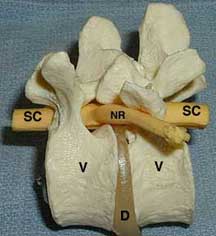
This side view of a spinal cord model shows 2 vertebrae (V) with a normal disk (D) in between. One of the nerve roots (NR) can be seen coming off of the spinal cord (SC). The head is towards the left, the tail towards the right.

To keep you oriented, this is the same area on a radiograph (at L1-2). The nerve root comes out of the dark structure that looks like a horse’s head. The disk, nerve root, and spinal cord do not show up normally on a radiograph. If the disk material becomes calcified it might show up.
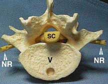
This view of a spinal cord model is an end-on view of how the spinal cord fits into the spinal canal. You can see how the spinal cord is enclosed by bone. If it swells it has no place to expand into, resulting in serious damage to the cord.
Pathophysiology of IVD
Classification
Type I occurs commonly in chondrodystrophic (poor cartilage and long bone development) breeds starting as early as 4 months of age.
The disk loses its moisture content and starts to mineralize. The stresses of daily living, especially jumping up and down, cause it to degenerate, ultimately rupturing its contents into the spinal canal and putting pressure on the nerve roots and spinal cord.
The pressure can be so great that the blood supply to the spinal cord can be damaged also, leading to actual death of spinal cord tissue (myelomalacia). Once this starts there is no treatment, and these dogs will become paralyzed and die of respiratory failure.
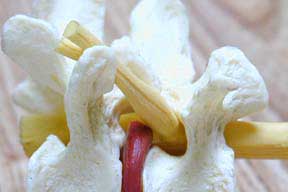
In IVD disease the disk material (red) in the space between the vertebral bodies puts pressure on the spinal cord and the nerve root that is leaving the spinal cord.
Type II occurs in larger dogs starting around 5 years of age. The changes in the disk occur much slower than in Type I disk disease. The disk bulges but does not actually rupture into the spinal canal. The spinal cord is not as severely injured and usually maintains its normal function. These dogs tend to show chronic pain and paresis.
The above classifications are helping in understanding IVD, but they do not always apply in every case. Large breed non-chondrodystrophic dogs can get Type I disk disease, and chondrodystrophic breeds can get Type II disk disease.
Degree of Damage
It is important to note that the speed at which a ruptured disk extrudes its material into the spinal cord is equally as important as how compressed the spinal cord becomes. Some dogs with minimal compression of the spinal cord can have severe neurologic problems because disk material extruded rapidly and severely damaged the spinal cord.
Cause
Trauma to a normal disk can cause disk material to extrude into the spinal canal. The most common cause is the natural degeneration of the disk that occurs in the chondrodystrophic breeds when young, and the larger breed dogs as they age. In most chondrodystrophic breeds it is the thoracolumbar area, the junction of the last few thoracic vertebrae and the first few lumbar vertebrae, that are involved.
Symptoms
The symptoms that occur vary from mild to severe. Much of it depends on which vertebrae is involved, how long the problem has been present, and whether the problem is Type I or Type II. It is important to follow the diagnostic process carefully when making a diagnosis.
The cervical (neck) vertebrae tend to have larger spinal canals for the spinal cord to pass through than do the vertebrae of the mid and lower back. When a disk puts pressure (whether Type I or Type I) the spinal canal has more room, so the spinal cord is subject to less compression in the neck than in the mid and lower back. Less compression means there is less of a chance that paresis or paraplegia will occur.
The following symptoms tend to occur with cervical disk disease:
- Crying- especially when the neck is manipulated or when lowered to eat or drink
- Poor appetite (anorexia)- pain can sometimes be so severe as to interfere with appetite
- Muscle spasms and reluctance to move- another sequelae to the pain that can occur
- One or both front legs might be lame- nerves to the front legs come out of the spinal canal at the cervical vertebrae.
- Ataxia- pressure on the spinal canal at the cervical vertebrae can interfere with the nerves that innervate all 4 legs. Only rarely will this cause tetraplegia (paralysis).
When IVD occurs at the junction of the thoracic and lumbar (thoracolumbar) vertebrae, symptoms might be different than in the cervical version. Some of these symptoms depend on whether there is a Type I or Type II problem.
- Crying in pain or shaking- a consistent symptom noticed by owners is their dog crying as if something hurts. It might happen spontaneously, or it might happen when you pet or pick your dog up. Those of us that have had a pinched nerve understand how severe this pain can become.
- Reluctance to move- this might manifest itself as a hesitation to jump onto the bed, reluctance to go up or down stairs, or just laying around more than usual.
- Poor appetite (anorexia)- the pain that occurs can decrease the appetite.
- Ataxia to rear quarters- a dog might walk around as if the back end is going in a different direction than the front end. This is caused by pressure on the nerve roots that go to the rear legs.
- Paraparesis or paralysis to rear legs- the pressure on the nerve root can become so severe that it can completely impair the nerve and cause paralysis.
- Tense abdomen- this is called referred pain, and can mimic the symptoms of other diseases.
- Hunched appearance- an additional problem related to pain
- Fecal or urinary incontinence- these are relatively severe signs of thoracolumbar disease
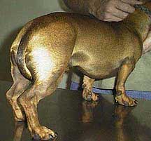
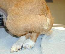
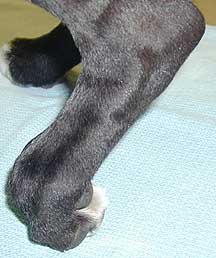
This dachshund is exhibiting signs of pain to its rear quarters. His tail is between his legs and his abdomen is hunched up.
This dog is “down” in his rear quarters, a potential sign of IVD disease
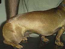
This dog is exhibiting serious signs of IVD. It is partially paralyzed in its rear legs.
This dog is paralyzed in its back legs
When the problem progresses to this point these dogs commonly will not be able to urinate. The urine needs to be manually expressed before it passively overflows. Urine that stagnates in the bladder causes discomfort, stretches the muscles in the bladder wall so much so that muscle tone will not return, and sets up the stage for a bladder and kidney infection.
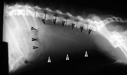
The arrows outline the distended urinary bladder of a dog with this problem
Diagnosis
Since the symptoms of IVD disease mimic those of other diseases, a thorough approach is needed to differentiate them. In every disease we encounter we follow the tenet’s of the diagnostic approach to ensure that we make an accurate diagnosis and that we do not overlook some of the diseases that are also encountered in pets as they age.
Signalment
IVD disease can occur at any age, although it tends to be a problem that affects middle aged and older dogs.
Several canine breeds are prone to getting IVD. They are called chondrodystrophic due to the shape of their bones from breeding:
- Dachshunds (most common breed by far- up to 18% will get this disease)
- Welsh corgi
- Lhasa apso’s
- Shih Tzu’s
- Cocker Spaniels
- Bulldogs
- Beagles
- Pekingese
History
IVD disease is suspected in any pet that has some of the symptoms described above, especially if the dog seems in pain or has some degree of paralysis. Falling or being hit by a car gives us a clue that the spinal cord might be damaged.
Physical Exam
Routine physical exam findings might include:
- Shaking
- Increased heart and respiratory rates
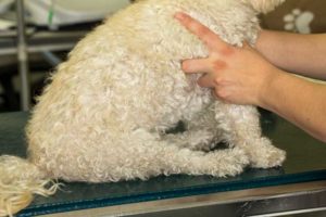
- Pain upon abdominal palpation
- Pain when moving the neck
- Weakness to the back end
- Lameness to any of the legs
During your dog’s exam you will notice one of our doctors checking some reflexes. This exam helps localize the problem and helps to verify that the problem is indeed IVD and not some other problem. Four of the more common neurologic tests will be explained:
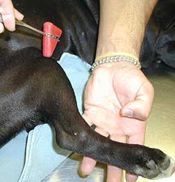
Panniculus
This test is performed by poking the skin gently with a needle. If the pin is felt the muscles underneath will cause the skin to temporarily “crawl”. By finding the junction where the skin no longer crawls it is possible to help localize the nerve root (remember, they overlap). The affected nerve root is usually 1-2 vertebrae in front of the spot where the skin crawls.
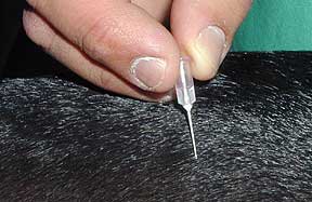
A gentle poke with a tiny needle is all we need for this test
Proprioceptive deficits
This is a postural reflex that tests the ability of a dog to recognize the placement of one of its limbs without actually seeing it. This tests the ability of the nerve to respond to the abnormal sensation to a foot that is bearing weight on the wrong surface.
When the wrong surface is bearing this weight it sends a signal from the nerves in the top of the foot to the spinal cord segment innervated by that foot. When the signal gets to the spinal cord segment it also travels to the brain signaling that the foot is in an abnormal position. The brain then sends a signal back down the spinal cord to the muscles that innervate the foot telling them to contract and put the foot back into normal position.
This is a sensitive test, and if a dog does not return its foot to a normal position immediately it potentially indicates a serious problem with the spinal cord. It is easily performed by placing the foot in an abnormal position. This dog should have returned its foot to a normal position immediately. Other diseases besides IVD can cause this problem (ex.- a fractured leg)
If the foot stays like this there is a deficit
Knee jerk (myostatic)reflex
This is a spinal reflex that tests the ability of the spinal cord to react to stretching of the patellar tendon. If absent or diminished it indicates a potential problem with the nerve root. If exaggerated it indicates a potential problem with the spinal cord. Other tendons besides the knee can be checked for this reflex.
Every pet has a different response to this stimulus, so it is usually repeated for accuracy
Deep pain (withdrawal) reflex
In this reflex a toe is pinched, which normally will result in withdrawal of the limb away from this painful stimulus. This occurs independent of whether or not the pain is perceived at the brain level (you already learned this at the beginning of this page in the spinal cord anatomy section). A pet with a problem in its spinal cord will have the reflex, but will not realize it is painful because the nerves that travel along the spinal cord to the brain are injured.
Dogs that show no reaction when a painful stimulus is applied to their legs are considered to have severe spinal cord injury. These dogs carry a poor prognosis for recovery, and most need immediate surgery. This is a subjective test though, and needs to be performed numerous times for proper interpretation. Some dogs don’t consciously show signs of pain, so this critical test can be misinterpreted.
Diagnostic Tests
Radiography
In some cases a radiograph is the diagnostic test of choice. Radiographs help determine if a dog’s pain or paralysis is due to IVD or some other cause. Other causes can include trauma, tumors, cysts, or infections of the vertebrae.
Dogs that have IVD might have calcified disks, collapsed disks, even calcified disk material in the spinal canal. Dogs that are radiographed for IVD disease must be under sedated or under general anesthesia for proper technique and positioning.
Some dogs with this arthritis are in pain and need medication, while others have no symptoms at all. Cats can get spondylosis also, although it is more common in dogs.
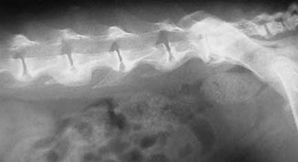
This dog has spondylosis, which is a form of arthritis due to instability of the vertebrae. This is not necessarily IVD disease.
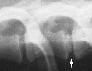
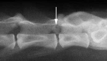
This radiograph of a Pug shows a collapsed disk along with spondylosis between L- 2 and L- 3. You can tell it is collapsed when you compare it to the width of the disk in front and behind.
This dog has IVD disease. There is calcified disk material in the spinal canal between L-2 and L-3, and it is painful and weak in its rear legs. It has a funny name- joint mouse.
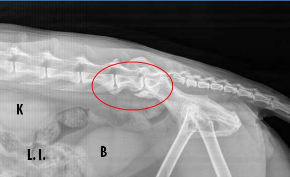
This is a cat with the problem in its last two lumbar vertebrae.
K-Kidney
L.I. – Large Intestine
B- Urinary Bladder
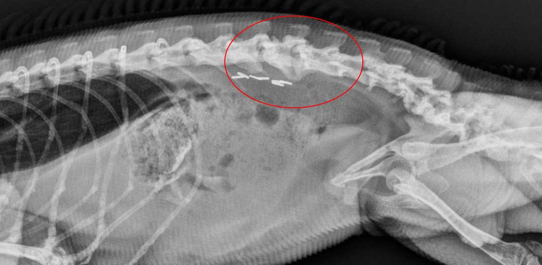
Spondylosis also occurs in animals like this iguana. The spondylosis is circled in red, along with metal clips, called hemoclips, used to tie off the blood supply to the ovaries when it was spayed.
This radiograph is from an 11 year old dog that is weak on one of its rear legs. Foreign bodies and infections are possible causes, but most likely it is a tumor. What the owner that was a “tweaked back” on their pet turned out to be a cancerous tumor, emphasizing the importance of radiography in diagnosis.
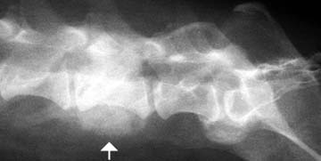
The arrow points to the involved vertebrae.
This is the same dog but from the VD view
Myelogram
As good as an x-ray is in making this diagnosis, it does not give all the information needed in some cases. Plain x-rays do not allow visualization of the actual spinal cord or nerve roots. The dye injected during a myelogram outlines the spinal cord and allows much better visualization of any pathology in the cord. Injecting a radiopaque dye into the spinal canal is of critical importance if surgery is contemplated.
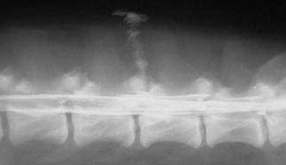
This myelogram is outlining the spinal cord in the lumbar vertebrae of a dog. The vertical column of dye at the top is where the needle was inserted to inject the dye. Now we can actually visualize the spinal cord itself.
Magnetic Resonance Imaging (MRI)
This tool is available to veterinarians and is very valuable in making a diagnosis of spinal cord disease. If the myelogram is inconclusive the MRI can provide valuable information on the health of the spinal cord.
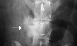
Cats can get spinal lesions also. This is the radiograph of a 15 year old cat with a lesion in its spinal canal at the arrow. This cat is painful and not walking well on its rear quarters.
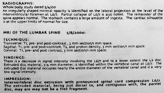
This is the x-ray and MRI report on this same cat
Treatment
Treatment depends on the severity of the problem and whether the problem is Type I or Type II. Fortunately, in many cases, especially if caught early, conservative therapy can be beneficial. Pets that have recurring problems might eventually need surgery.
Cage Rest
This is the most important treatment modality short of surgery. Cage rest means exactly what is says; there can be no running, jumping, or playing. A crib or playpen will not work because dogs will try to jump out. It sometimes has to be utilized for several weeks for an adequate outcome.
Dogs that are cage rested need to be monitored carefully for progression of the problem. Serial neurologic exams are used to monitor their condition. They should be hospitalized for the first few days in order to monitor their progress.
Medication
Anti-inflammatory and analgesic medications are used routinely in Type I disease. Medications are highly beneficial in reducing pain and minimizing inflammation at the delicate spinal cord. The mainstay when it comes to medication is cortisone.
Muscle relaxants are also used to minimize the spasms that accompany this problem. It is imperative that any pet with IVD that is put on medication is under strict cage rest.
Pets that feel better with medication might be inclined to resume their normal activity, greatly increasing the chance that their spinal cord will suffer more damage.
Our goal is to give just enough pain medication to make the dog more comfortable, yet not so much that the dog resumes its normal activity. If a dog’s condition improves with cage rest it should be continued for up to 3 weeks. Exercise should be restricted for an additional 3 weeks.
Unfortunately, recurrence is common. Dogs that are unable to urinate properly due to this disease might also be put on antibiotics to prevent urinary tract infections.
Dogs with Type II are routinely placed on anti-inflammatory medication. There are many highly effective ones available to veterinarians that greatly enhance a dog’s quality of life. You can learn more about these medications, called NSAID’s (Non steroidal anti inflammatories) in the treatment section of our arthritis page.
Surgery
Surgery is the treatment of choice for recurring problems, dogs that have not improved with conservative therapy, or those that have neurologic deficits. Dogs that are paralyzed (paraplegic) in the back legs need immediate surgery. During the surgery the goal is to relieve the pressure on the spinal cord. This is done by removing a piece of the vertebral body or cleaning out the disk material that is putting pressure on the cord.
Surgery is also used to verify a diagnosis since the actual spinal cord can be visualized. In addition, even if the prognosis for recovery from paralysis is poor, surgery can minimize pain at the spinal cord if an owner is willing to deal with the long term paralysis.
Post operative care is important in dogs undergoing back surgery. They might need hydrotherapy, manual expression of their bladders, controlled walking with assistance, and lots of TLC.
This chart gives an idea of how these treatment modalities are used. These are not hard and fast rules, but more of a guideline.
| Medical Therapy | Surgical Therapy |
| First episode of pain only | Several episodes of pain only |
| First episode of paresis or mild ataxia | Several episodes of paresis or mild ataxia |
| Medical conditions that prevent surgery | Condition that worsens with only medical therapy |
| Paralyzed and no deep pain response for > 48 hours | Mild paralysis with deep pain response present |
| Symptoms of progressing myelomalacia | Paralyzed and deep pain response present for less than 48 hours |
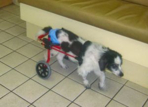
A paralyzed dog can lead a very high quality of life. These dog carts work quite well.
Acupuncture
A relatively new treatment modality for IVD in dogs is acupuncture. It can be helpful in dogs that are not paralyzed or in those where anesthesia or surgery are contraindicated. Our acupuncture page has more details.
It is important to remember that it does not always work, and the prolongation of other treatment modalities should not be undertaken due to the severe and potentially irreversible nature of this disease.
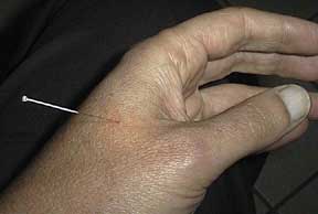
Dr. P wanted to make sure the needle did not hurt an already painful dog, so he volunteered to be the “pain guinea pig”. He is happy to report that there was no problem (and he could now actually dunk a basketball!).

Here are the needles in use in Addison (the Pug whose x-rays you saw previously).
VNA (also called VOM)
In the last few years our doctors have been using this treatment method in almost every case of IVD when the pet is not paralyzed. It is called VNA, and it stands for Veterinary Neuronal Adjustment. It has changed the way we look at this disease, and give us a new tool to treat without the use of drugs. Our VNA page has more details.
VNA is used on a variety of different species like this iguana
We even use it on rabbits
In addition to VNA we also use the Therapy Laser as mentioned in the dog VNA video. Our Therapy Laser page has the scoop on this treatment of this painful disease also.
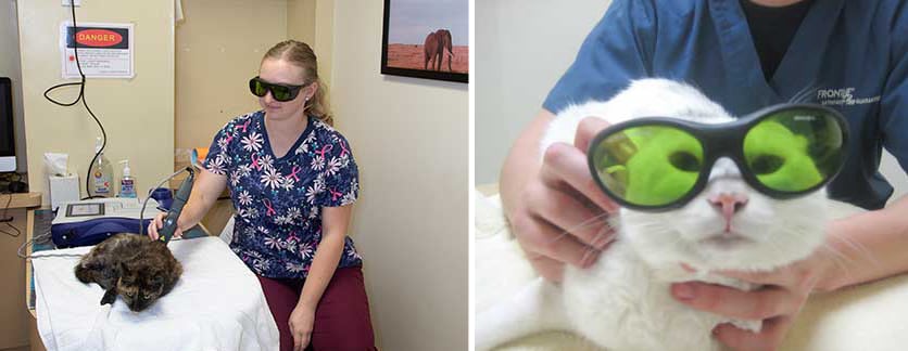
Pets that are uncomfortable or painful from disk disk just lay there while we treat them. They even get to wear the cool laser glasses if they want!
Prevention
Since this seems to be a heritable disease it is theorized that screening of dachshunds at 2 years of age with radiographs, looking for disk calcification, can be an early indicator for IVD.
Keeping these dogs out of the breeding pool might decrease the incidence of this disease. This is similar to taking radiographs for hip dysplasia at 2 years of age to screen for this problem before breeding.
Keep your dogs weight normal for its breed, and do not let it do any excessive jumping.
Be observant for any signs of this problem so we can institute medical therapy and not need to resort to surgery.
Prognosis
IVD is a serious and potentially crippling disease. If your dog exhibits any of the previously described symptoms it needs an immediate exam by one of our doctors. If the problem is caught early enough the outcome of this disease is usually satisfactory. If you own one of the breeds that is predisposed to this disease it is important to closely observe for the symptoms of IVD disease.