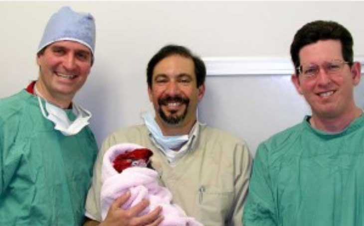
With the significant help of our favorite veterinary ophthalmologist, the late Dr. Paul Jackson, along with our favorite human ophthalmologist, Dr Art Giebel, we removed a cataract from a species of bird called a Macaw.
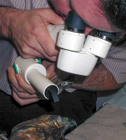
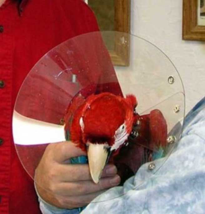
Paul helping us with a hawk from our Wildlife Program that has an injured eye
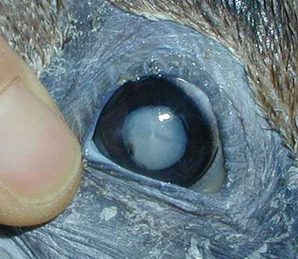
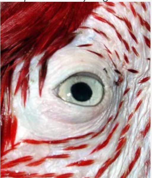
Even wild birds like this pelican get cataracts
We now work closely with Doug Esson at Veterinary Ophthalmic Consulting in Irvine, CA for our avian eye problems.
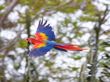
Before we get on to this surgery, we would like to show you a picture of a beautiful wild Macaw that Dr. P took on a recent trip to Costa Rica. If you click on the photo you can learn more about how he teaches wildlife photography around the world.
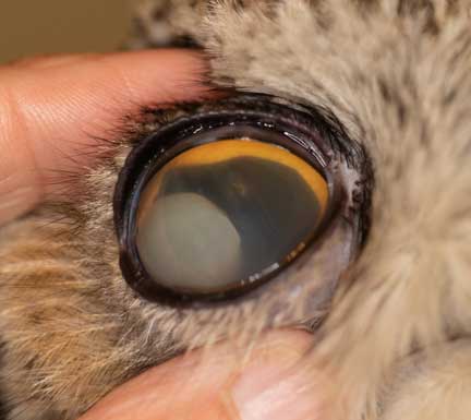
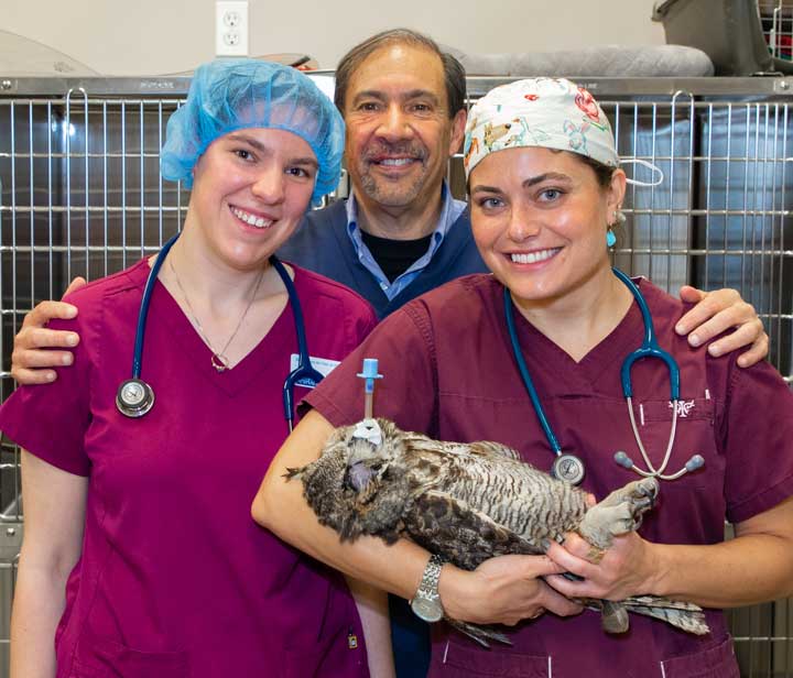
This is not a routine surgery, and requires the skills of a veterinary ophthalmologist like Dr. Jackson. At the end of this page you can see how another veterinary ophthalmologist named Doug Esson of Veterinary Ophthalmic Consulting (VOC) helped us with an owl with an eye problem that was from our Wildlife Program.
This page has graphic surgical pictures.
Pre-anesthetic Preparation
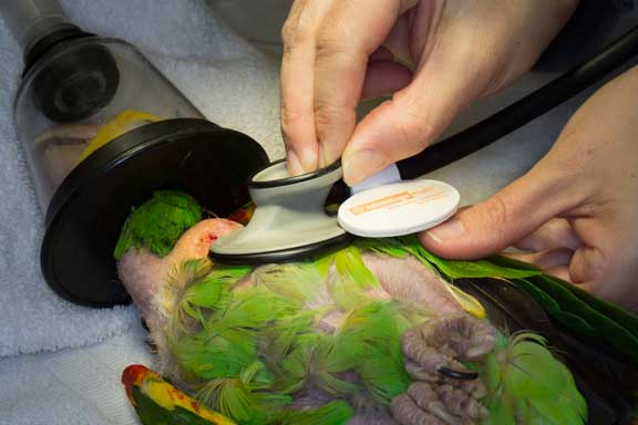
The first step in the process is a thorough physical exam. This is performed immediately prior to surgery.
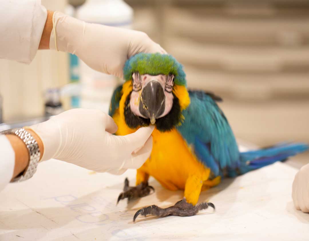
This young Macaw is enjoying the attention he is getting during his exam
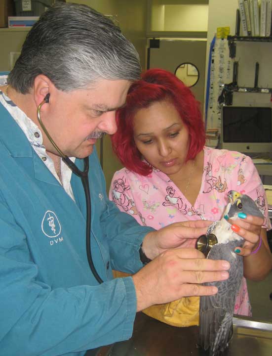
No matter what type of bird, all of them are given a thorough exam prior to surgery. This is a peregrine falcon from our Wildlife Program.
We also perform pre-anesthetic diagnostic tests prior to surgery to make sure there are no internal problems.
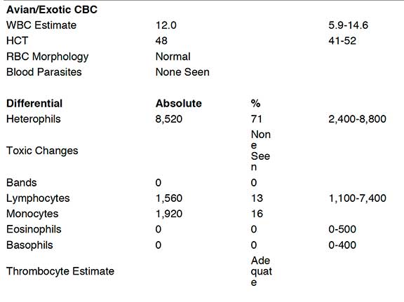
This part of the pre-anesthetic blood panel checks the red and white blood cells. It is called a CBC
Anesthesia
Birds tend to be more sensitive to anesthesia than most mammals, so special precautions are taken to minimize the risk.
We use general anesthesia because this is delicate surgery and there can be no movement of our patient

Equipment
In addition to the substantial surgical expertise of our surgeons, this surgery is not possible without special equipment, especially a dual surgical microscope.

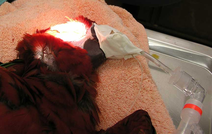
Our patient is anesthetized and ready for surgery
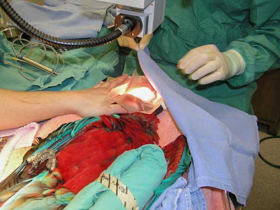

Dr. Paul and Dr. Art work together as a team during the surgery
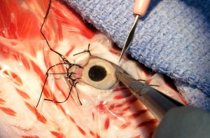
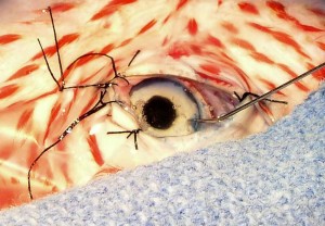
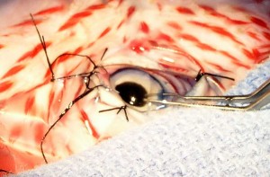
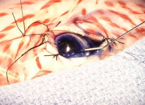
Surgery
A microscopic incision is literally made into the eye near the cornea. The cataract is emulsified just like in people. A new lens is not put in, unlike in people.


Art Giebel, MD
Carl Palazzolo, DVM