
Introduction
Tumors of the adrenal gland in ferrets can cause excess secretion of sex hormones, thus affecting many organs in the body. Unfortunately, this is a relatively common problem in middle aged and older ferrets, although it can happen at any age. Even though most of these tumors are not malignant, they can cause disease if left untreated.
This disease goes by several names in addition to the one in the title of this page:
- ADG- adrenal gland disease
- AGN- adrenal gland neoplasia
- ACD- adrenal cortical disease
- AEE- adrenal associated endocrinopathy
- FADC- ferret adrenal disease complex
FADC is probably the most accurate description, because as is the case with so many diseases, as we advance our knowledge over the decades, and develop more sophisticated diagnostic tools, we realize that most diseases are much more complex than originally envisioned. It is human nature to try to make things simple so they are easier to understand. This does not apply to most diseases we encounter in animals.
Dogs and cats get a problem similar to this, although it acts and is treated differently. In dogs and cats it is due to an excess secretion of cortisone, not sex hormones. In these species it is called Cushing’s disease.
We are a teaching hospital registered with the University of California at Davis (and most every veterinary school in the country and many overseas veterinary schools), so you will see how we perform this vital function for veterinary students and new graduates in this page.
These students post a Daily Diary of what they learned at the Long Beach Animal Hospital each day of their externship. This one is from a veterinary student in her senior year at the University of California at Davis. You can see their Facebook posts on our Facebook page.

At the end of this page is a video of part of a surgery to remove cancerous adrenal glands and cancerous nodules on the pancreas.
Why Baby Ferrets Need Vaccines
Ferrets are highly susceptible to Canine Distemper, and if they get it the disease is usually fatal, so vaccines are imperative. We like to give them at 8, 11, and 14 weeks of age. We also give them rabies vaccines, although we prefer to space them apart from the Distemper vaccines.

We couldn’t resist adding them to our page they are so cute!
What Causes Adrenal Disease in Ferrets
The exact reason this tumor arises is not completely known. It is seen more often in the U.S. than in Great Britain, where different breeding and husbandry practices are utilized. It is speculated that diet, exposure to sunlight, and neutering are all factors, with neutering being the most important one.
Ferrets breed seasonally, causing variation in melatonin release with varying daylight. Less daylight means more melatonin and a thick haircoat. This higher level of melatonin eventually exerts a negative feedback on the release of the sex hormones estrogen and testosterone. When ferrets are spayed and neutered the negative feedback is disrupted, more of these sex hormones are secreted than is normal, and clinical signs develop.
Types of Ferret Adrenal Gland Tumors
The three main types of adrenal lesion encountered are:
- Benign nodular hyperplasia that occurs 56% of the time
- Benign adrenocortical adenoma that occurs 16 % of the time
- Malignant adrenocortical adenocarcinoma that occurs 26% of the time
- A combination of the above
What is Normal Ferret Adrenal Physiology?
This disease involves reproductive hormones. In a normal ferret, a hormone from the hypothalmus in the brain, called gonadotropin-releasing hormone (GnRH), is released in larger amounts, usually due to an increase in daylight. This causes stimulation of luteinizing hormone (LH) and follicle stimulating hormone (FSH) from the pituitary gland.
These hormones stimulate the release of estrogen and testosterone from the gonads and adrenal glands (important point in neutered pets that have no gonads). A very sensitive negative feedback loop maintains just the right amount of estrogen and testosterone. This sensitive balance is upset in adrenal disease of ferrets.
In the medical treatment section we will discuss a drug called Lupron and Deslorelin. This drug binds with receptors on the GnRH molecules and lessens its effects on the pituitary.
Ferret Adrenal Anatomy
The adrenal glands are small glands located just in front of the kidneys. The left gland is embedded in fat just in front of the kidney, the right one is located deeper in the abdomen and under one of the liver lobes. The left adrenal gland is the one affected in almost all cases, which is advantageous surgically as you will learn.

The arrow points to a normal left adrenal gland of a ferret. It is quite small and buried in fat in front of the kidneys. Click on the photo to enlarge.
A close up to shows how tiny it can be when normal

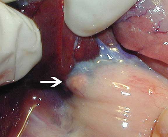
Here is another one at the top of the hemostat on the left. This one is inflamed due to adrenal disease.
The tip of this hemostat shows an inflamed adrenal gland. Note the large horizontal and blue vein underneath it, called the adrenolumbar vein. This is the vein that will be ligated with metal hemoclips you will see later when we show surgery removing the adrenal gland.

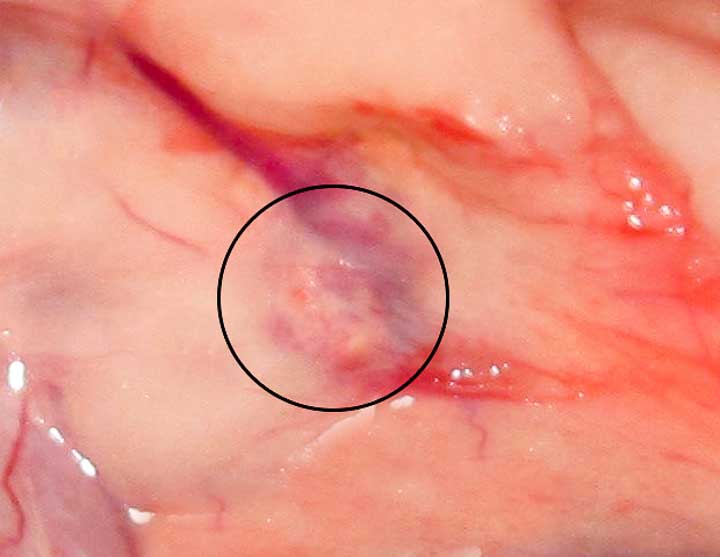
The left adrenal gland to the right of the arrow is very inflamed

The left adrenal gland to the right of the arrow has even more inflammation

The right adrenal gland is small, and under the right lobe of the liver, adjacent or attached to the vena cava (VC). The vena cava is the major blood vessel for the return of all the blood from the back of the body to the heart. The liver has to be pulled away to visualize the right adrenal gland, which is why you cannot see the adrenal gland in this photo.
When you pull the liver lobe forward you can see the enlarged right adrenal gland to the right of the arrow. You can also see how the right adrenal gland is adhered to the vena cava (the blue horizontal structure), which makes surgery to remove a diseased right adrenal gland problematic, to say the least.

A different view of the right adrenal gland and its close association to the liver and vena cava (VC)

The white arrow in the lower left of this picture points to a normal right adrenal gland. It is very small because it is normal. On the left, above this right adrenal gland, is a lobe of the liver (L) that has to be pulled forward during surgery in order to get access to the right adrenal gland. The posterior vena cava is the large blue vein running horizontally. The abnormal left adrenal gland can be visualized to the left of the arrow on the top right. It will be removed.
What Happens When Adrenal Disease is not Treated?
When a diseased adrenal gland is left untreated it might eventually get extremely large and cause substantial illness. Symptoms of adrenal disease would usually appear long before the gland gets this large, and it would hopefully be surgically removed before getting this big.

Sometimes the tumor on the right side gets greatly enlarged, and be as big as the kidney. Here it is intertwined with the right kidney and the vena cava (vertical blue line to the right of the tumor). This would be very difficult to remove surgically because of the involvement of the tumor with the vena cava.

Here is another large adrenal tumor (T) under a lobe of the liver (L)
What is the Pathophysiology of Ferret Adrenal Disease?
These small but very important glands have numerous roles you can learn more about in our Cushing’s page. It gets very detailed, so be prepared to learn some anatomy and physiology before you return back here to finish learning about adrenal disease in ferrets.
In Cushing’s, the adrenal glands secrete too much cortisol from a part of the adrenal gland called the Zona fasciculata. In Adrenal Disease in ferrets, it is the Zona reticularis, the area that secretes the sex hormones estrogen and testosterone, that is stimulated to produce too much of these hormones.
Here is a breakdown of the important sex hormones secreted by the adrenal glands:
- 17 – OHP (17 alpha hydroxyprogesterone)
- androstenedione
- DHEa (dehydroepiandrosterone sulfate)
It is postulated that these hormones are secreted from the adrenal glands after chronic stimulation from follicle-stimulating hormone (FSH) and luteinizing hormone (LH) from the pituitary gland. Indoor housing of ferrets, leading to more light than ferrets in the wild, is postulated to also stimulate release of LH, along with gonadotropin releasing hormone (GnRH) from the pituitary gland.
The end result of this hormone imbalance is excess secretion of estrogen in females and testosterone in males. This causes the vulva to enlarge in females, and a return to male sexual behavior in the male. The enlarged vulva is easy to see, which might be the reason we diagnose this problem more often in female ferrets.
A special lab in Tennessee can check for excess amounts of these hormones on a blood panel, an important help in the diagnosis of this disease. A ferret can have normal values on this test, yet still have adrenal gland disease. This is why we always follow the tenets of the diagnostic process.
What Are the Symptoms of Ferret Adrenal Disease
Hair loss
The most common symptom of adrenal disease in ferrets is hair loss, sometimes with itchiness (pruritis). Itchiness due to adrenal disease can also be present with no hair loss. The hair loss can be seasonal, can come and go, but eventually it progresses to almost complete baldness. The most likely spots for hair loss are the tail area and rear legs. Hair loss in dogs and cats is almost always caused by a different problem. This hair loss occurs in over 80% of the ferrets with this disease.
Do you see the hair loss at the back end of this ferret?

This view gives you a better idea of the extent of the hair loss

She even has a bare tummy

At least she is not as bad as this ferret

Hair loss can be anywhere on the body. This is typical of the hair loss that can occur at the tail

If you look at only the tail it is hard to tell if it is a ferret or a rat!
Enlarged Vulva
Sometimes we see enlarged vulvas in spayed females. This can be difficult to see in the early stages due to the small size and anatomic location for an animal that is always low to the ground.

This one is slightly swollen. It should not be swollen at all since she is spayed.

This one has more enlargement. Notice the hair loss?
A female ferret can have an enlarged vulva because it is in heat (very rare since all of them are spayed prior to purchase) or because a remnant of ovary is left after the surgery. Since most ferrets do not develop adrenal disease until 3 years of age or more, an intact female ferret, that goes into heat at between eight and 12 months as evidenced by a swollen vulva, usually does not have adrenal disease.
Difficulty urinating (stranguria) in the male ferret
Male ferrets sometimes have difficulty urinating in addition to hair loss. Even if they are neutered there might be a return to normal sexual behavior. There might also be lethargy, a decrease in appetite and weight loss. Other symptoms include urinating outside of a litter box, aggressive behavior, or general discomfort.
In male ferrets cysts can occur in the small amount of prostatic tissue they have. They can also get a bacterial prostatitis causing these symptoms. This can cause difficulty in urination, which can affect the kidneys if it progresses. On rare occasion this problem can progress to complete inability to urinate due to the enlarged prostatic tissue. Inability to urinate is a medical emergency, and needs immediate catheterization through the urethra to save the kidneys and prevent the urinary bladder from rupturing.
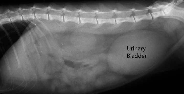
A urinary bladder that is so distended that a pet cannot urinate is diagnosed on palpation during a physical exam. It is confirmed with a radiograph, as in this cat that cannot urinate. If we run a blood panel on this animal we will see what is called post renal azotemia, a sign the kidneys are shutting down. Our kidney page has much more detail on this.

This is the necropsy picture of a severely distened bladder in a ferret that did not make it

Let’s learn our normal ferret urogenital anatomy first. Circled in red is the os penis, which is a bone. This is the same as a dog, except note how the tip of the os penis curves upwards.

The os penis opening is very small, and the tip is curved, making catheterization difficult

The leftmost red circle shows the kidneys, while the rightmost red circle show the tip of a urinary catheter in the urinary bladder, allowing urine to flow out of the bladder. You can see where this urinary catheter started by looking at the tip of the os penis, and then follow it around (the pelvic urethra) until it ends in the urinary bladder.
The diameter of the urethra is small, and it is lined with cells that are easily traumatized and can be irreversibly damaged by a catheter. Add the fact that the os penis prevents the urethra from expanding, and you have potential for complications while passing the catheter. Then add the fact that the tip of the os penis is curved, making initial entry of the catheter problematic. This is why an experienced veterinarian is needed to put this catheter in. This is also why a ferret is commonly anesthetized for this procedure.

A radiopaque dye can be injected into the bladder to make sure it has not ruptured. A rupture is a medial emergency and requires immediate surgery if this ferret is to live.
Urethral and Cystic (urinary bladder) calculi
Stones in the urethra and bladder can also cause stranguria, so we cannot always assume adrenal disease is the cause of a ferret that is straining to urinate or cannot urinate.

These are what stones look like in a ferret urethra
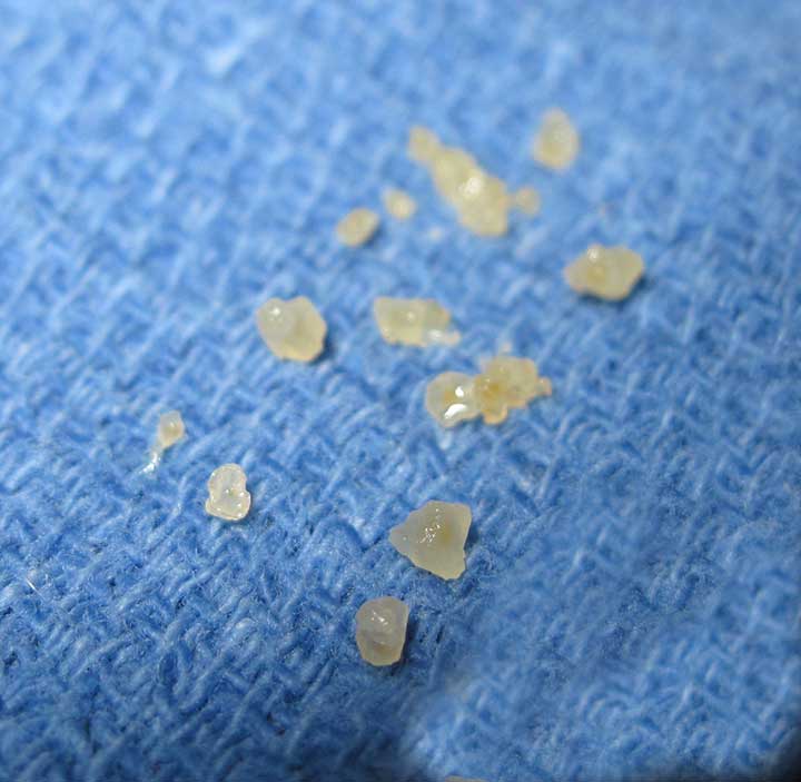
The same stones after removal from the urethra
Male ferrets with adrenal disease might also return to typical male sexual behavior or mounting, whether it is a female or a male they are mounting. Aggression can occur, and seems to occur more in ferrets with malignant adrenal disease (adenocarcinoma).
Anemia
Almost all ferrets in the United States come from a company called Marshall Farms. They routinely spay, neuter, and descent all ferrets before distributing them to pet stores and other dealers.
In years past, before all female ferrets (called Jills) were routinely spayed, an enlarged vulva in a sick ferret was a sign of a ferret that did not mate when it was in heat. Being an induced ovulator, meaning Jills would only release eggs from their ovaries when they mated, Jills that did not mate would secrete an excess of estrogen from their ovaries for a long period of time.
This excess estrogen was toxic to the bone marrow, and could cause a severe life-threatening anemia. The technical term was aplastic anemia. The anemia would show up as severely low RBC’s (red blood cells), HGB (hemoglobin) or HCT (hematocrit), and even thrombocytes (clotting factors) would be low.

This ferret has a hematocrit (HCT) of 15.5 %, typical of a ferret with aplastic anemia. It should be at least 43%. This is severe and would require a blood transfusion to keep it alive.
On very rare occasions the adrenal gland tumor (instead of the ovaries this time) can secrete estrogen, which in the ferret causes a severe anemia as you already learned. This ferret might need a blood transfusion prior to surgery to remove the adrenal gland.
Lethargy and muscle atrophy
Occasional symptoms might include lethargy and muscle atrophy. Abdominal muscle atrophy can also cause the pot-bellied appearance seen in Cushing’s disease of dogs.

Sometimes the muscle atrophy is severe, and is also caused in conjunction with other common ferret diseases, notably cancer in the intestines or lymph nodes. The bulge on the left at the back end of this ferret is the right kidney.
How do we Diagnose Ferret Adrenal Disease?
We always follow the tenets of the Diagnosis Process in making this diagnosis. In ferrets these are the typical findings:
Signalment
This tends to be a disease of neutered male and female ferrets with an average age of 3 years. It be seen earlier and later than these age groups.
Symptoms
Symptoms of adrenal disease in ferret were described earlier in this page.
Physical Exam
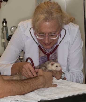
Ferrets are fun critters to work with medically. They can be a challenge though when trying to listen to their heart with a stethoscope or perform some other detailed exam, due to their non-stop movement and curiosity.

These guys are troublemakers, and are so fast and sneaky that before you know it one is up your sleeve before you can even start your exam!

They are curious though, so they will soon come out and look for somewhere else to cause trouble !
We perform a complete physical exam on all our patients no matter what disease we suspect is causing a problem. This is particularly important in ferrets due to their great propensity to get other diseases besides adrenal gland disease. This might reveal pale gums and heart murmur if there is a significant anemia. If the adrenal gland is on the left (most cases), and it is large enough, it might be possible to feel it during abdominal palpation.
Although enlarged adrenal glands can be palpated during an examination, this is not usually the case. A large number of adrenal tumors are confirmed during routine exploratory surgery, especially if the ferret exhibits the typical pattern of hair loss and an enlarged vulva in the female, or straining to urinate in the male. Exploratory surgery is a common way to verify the diagnosis and correct the problem. During this surgery we routinely check other organs for problems, especially the pancreas for tumor nodules that might be an insulinoma.
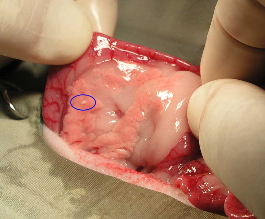
That little nodule circled in blue is an insulinoma tumor in the pancreas of a ferret during exploratory surgery to check for adrenal disease. You will see this adrenalectomy surgery later in this page.
Initial Treatment of an Ill Ferret
These ferrets can come in very ill, and need medical stabilizing before we can proceed further. At this point in the exam we do not know what disease an individual ferret has, so before we run some diagnostic tests we start correcting basic problems like dehydration, hypothermia, negative nitrogen balance, nausea, and hypoglycemia before proceeding further. This is important in such a small animal with a high metabolic rate that can easily go hypothermic (low body temperature) and hypoglycemic (low blood sugar) on presentation.
This ill ferret needs immediate medical treatment

Since they are highly prone to hypothermia we need to immediately know the body temperature

The same thing holds true for its blood sugar (glucose) level

We train our staff and students that gentle handling is a must at all times
Treatment with Intravenous (IV) Fluids
IV fluids are a crucial part of our medical treatment in all species for a wide variety of reasons in a vast array of diseases. It is so important we have a page dedicated to the medical use of fluids in animals. Click here to learn more, but don’t forget to come back here because we have much more to teach you about adrenal disease in ferrets.

Most ill ferrets are treated with IV fluids to correct dehydration. The IV catheter also allows easy administration of medication without having to inject into the muscle, along with anesthetics and sedation.
Diagnostic Tests
Routine Ferret Blood Panels
Our lab can run a series of sophisticated tests on the small amount of blood we get from a ferret. We routinely run this blood panel on Wellness Exams for early awareness of health conditions. We also run it on all sick ferrets to help make a diagnosis when they are sick, and those that will soon have surgery to make sure the liver and kidney can handle anesthesia if we need to surgically remove an adrenal gland tumor.
It is especially important to run a blood panel to make sure we see no signs of cancer, which is common in ferrets, and We also carefully assess the blood sugar (glucose) level to give us any indications of insulinoma, another common ferret cancer.

This ferret has a low blood glucose level, and this finding warrants further investigation by checking an insulin level

The insulin level came back elevated at 47, so this pet needs further treatment that you can learn about in our insulinoma page

We also like to perform a urinalysis to assess the urinary bladder and kidneys
Now that all ferrets are spayed and neutered upon purchase in the United States, we no longer see this type of anemia. When we have a ferret with an enlarged vulva we know it is most likely due to adrenal disease, especially if hair loss is present also. Retained ovarian tissue form a spay can cause this on rare occasion.
Adrenal Panel
Sex hormone assays are very helpful in the diagnosis, particularly to rule out other diseases. You learned about these hormones in the physiology section above. This test can be used if the typical symptoms of hair loss and enlarged vulva or urine straining are not particularly prevalent.
We send all of our hormone tests on ferrets to the University of Tennessee.

This is their results based on 3,000 ferrets with confirmed adrenal disease, on how much of an increase (the abnormal column) is needed above the normal column to be considered positive for ferret adrenal disease. They also noted that all 3 tests need to be run because on occasion estradiol was normal in a ferret that has adrenal disease.

This is how they report a sample back to us. You can see the Estradiol is 254, way over the normal 180 of a neutered ferret.
This adrenal panel needs to be interpreted with regard to time of the year in the northern hemisphere, because even neutered ferrets can have seasonal variations in these hormones. This emphasizes the importance of the diagnostic process in making a diagnosis of this disease, and using clinical signs along with ultrasound, surgery, and biopsy findings, to confirm a diagnosis.
Urinalysis
If prostatic disease is present the urinalysis might show an infection. There might also be an elevated white blood cell called leukocytosis in the bloodstream of ferret with an infection.
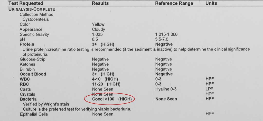
This urinalysis shows elevated white blood cells (WBC) along with a class of bacteria called cocci.
Radiology
Radiographs are usually unhelpful in this specific diagnosis since it is difficult to see the adrenal glands. On occasion, due to size or calcification, we can get an indication the adrenal glands are enlarged on a radiograph.

This normal ferret right lateral radiograph shows two overlapping kidneys (K) in the middle of the abdomen. The adrenal glands are located just to the left of these glands. They are not routinely visible on radiographs. See the labeled radiograph below.

The abbreviated organs are:
K- the two overlapping kidneys
LI- Large intestine
UB- Urinary blader
Can you see the the large whitish area on this radiograph that could indicate a greatly enlarged adrenal gland?
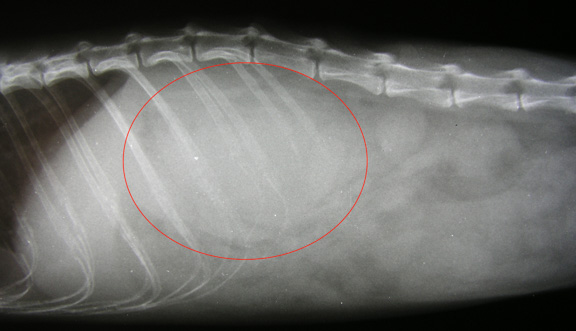
It has been circled for easier visualization
Radiographs are still indicated in a sick ferret due to the many other internal problems that can be diagnosed this way. As with many animals that hide their symptoms and do not talk to us about how they feel, you never know what you are going to find cooking inside of one…….

The arrow points to a circular foreign body with a hole in it that is inside of the small intestines

Here it is on another view

Our culprit in the small intestines trying to hide from us

This is what it was
Ultrasound
Ultrasound is what is needed to visualize the adrenal glands in ferrets, and also dogs and cats in most cases. Ferret adrenal glands can be tiny, which is why we always call our radiologist to perform this exam. Our radiologist, Dr. Ann Reed, is very experienced with ferrets, and is the only one we trust to perform our ultrasounds.

Most ferrets enjoy the attention and tummy rub as we gently move the probe on the abdomen

With ultrasound we can measure the size of the adrenal gland and determine if it is enlarged
Our ultrasound page has a nice explanation of this diagnostic modality that has greatly enhanced our ability to diagnose disease in animals. It has replaced exploratory surgery in many cases due it is accuracy and lack of invasiveness (the only invasive thing in an abdominal ultrasound is a tummy clip).
If you would like to learn more about diagnostic testing at LBAH follow this link.
Surgical Treatment of Ferret Adrenal Disease
The primary method of treating adrenal disease in ferrets is surgical removal of the gland. The problem can occur on either the right or left adrenal gland (or both). In the overwhelming number of cases the problem is in the left gland only. Eventually, both glands are commonly involved. After surgery to remove the affected gland vulvar swelling decreases and hair growth starts within 1-2 months and returns to normal at 6 months in most cases.
The left gland is much easier to work with because it is in a fat pad above the left kidney. The right gland is much more difficult to approach because it is under a lobe of the liver and is attached to the posterior vena cava, the main vein that returns blood from the back end of the body to the heart. You learned this in the pictures at the beginning of this page.
In most surgeries we remove the diseased left gland, leaving the right gland alone. Complete removal of both glands can cause serious complications. Sometimes removal of only one of the glands can cause a problem if the remaining gland cannot make up for the loss, and might lead to a disease called hypoadrenocorticism. This is rare in ferrets and not a reason to not remove one of the abnormal adrenal glands.
We take the same precautions in ferret adrenal gland surgery as we do in all pets. First we perform a pre anesthetic blood panel (you saw what it looks like earlier in this page) to check the internal organs. If everything is in order we perform a pre-anesthetic physical exam, and then carefully go through our checklist prior to anesthetizing our patient.
Dr. Kennedy has an assistant help hold a wiggly ferret for a pre-anesthetic exam

While our patient is brought into the surgical prep area our surgeons are getting ready
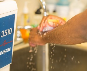
This is a sterile abdominal surgery, and our surgeon starts the pre-surgical process by using special soap to clean his hands
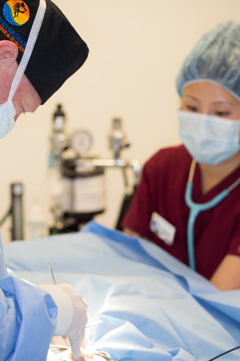
While our patient is being anesthetized our surgeon is already in our surgical suite setting up instruments. He is ready to start before our patient is at a proper plane of anesthesia. Once the anesthetist gives the green light the surgery starts immediately. We want our surgeon waiting for his patient, not the other way around, in order to minimize anesthetic time.

Only when everything is in order does our surgeon start the procedure
Time is of the essence in such a small patient in regards to anesthetic risk, low body temperature (hypothermia) and low blood sugar (hypoglycemia). Since we are a teaching hospital, our experienced surgeons, in this case Dr. Ridgeway, assists our more junior surgeons to make sure they operate efficiently and rapidly, and help handle any complications that arise.

Dr. R is helping Dr. Wood, while our extern watches and learns

Only once the surgery is almost complete does Dr. R leave the surgery for Dr. Wood to suture the abdominal incision
Our patients are carefully monitored to detect any abnormality before it becomes a problem. This early warning system is important in such a small animal that is ill and undergoing anesthesia and major surgery.

This machine monitors:
Temperature
Heart Rate
Heart rhythm
Oxygen saturation
Carbon dioxide level
Respiratory rate
We also carefully monitor oxygen saturation on our surgical patient with a special and small pulse oximeter
We also carefully monitor the blood pressure. These are normal numbers in a ferret under anesthesia. Note how fast the heart is beating (212 times per minute) in a small animal as compared to a person

In addition to our monitoring equipment our anesthetist stays “hands on” in monitoring important physiologic parameters. We use a special stethoscope (called an esophageal stethoscope) that is passed down the esophagus and can give us a clear sound of the heart.
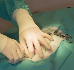
Once our patient is draped we are ready to proceed

We make our incision in a particular location in the center of the abdomen, called the ventral midline. We use a special scalpel made for a small animal like a ferret.

The next layer is a special muscle/tendon layer, called the line alba. It is important that we go through this particular area because this is the area that will be able to hold the sutures we use to sew the muscles back together, and thus prevent a hernia.

A special surgical scissors is used to extend the incision
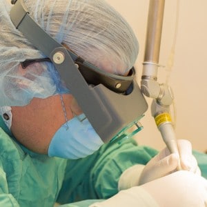
Ferrets are little guys, so their surgical anatomy is small. In some surgical cases we use magnifying loops to help identify structures.

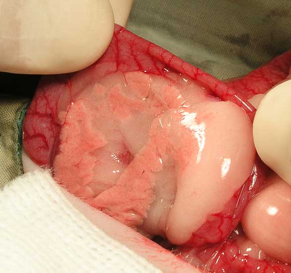
This picture is from an actual surgery. The inflamed left adrenal gland, buried in fat, is in the center of this picture
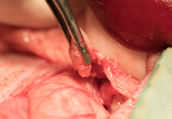
It is at the tip of this forceps after it has been dissected out of the fat

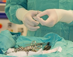
Special stainless steel clips, called hemoclips, are used to ligate the blood vessels as we remove the gland

This is the 1 cm diseased adrenal gland after removal, with some fat still around it
Use of the Surgical Laser in Ferret Adrenal Disease
We sometimes use the laser on the larger glands to aid in hemostasis (control of bleeding). This is of great important in such a small animal because large tumors can bleed excessively. Our laser has revolutionized some of the surgeries we perform on small animals like a ferret.

The tumor is on the right
While our patient is still anesthetized, although at a light plane since the surgery is over, we do several important things regarding pain to help post operative recovery:

We inject a long acting local anesthetic so our patient wakes up pain free

We also use the companion laser to decrease post operative pain and aid the healing process
Anesthesia
Since ferrets are prone to hypothermia, and even though we have been checking it continuously during surgery, we check it one more time before it is fully awake. You saw that earlier when we first perform an exam. This health parameter, along with many others, is continuously recorded during surgery.
You can see this in a small section of our anesthetic log below. All of these numbers are normal for a ferret under anesthesia, and the surgery was a success and the ferret woke up fine afterwards. There are several things to note on this log, starting with the left column we will go over the important ones:

This is only a small part of what is monitored and recorded during surgery
The surgery started at 2:50 PM and was completed at 3:35 PM, for a total of 45 minutes. We talked about the importance of getting this procedure performed rapidly to minimize anesthetic time and the complications that go along with this, now you are seeing this in actual numbers.
The next column shows the continual monitoring of the temperature, which in this case stayed normal the whole time. This is because we use external heating pads and blankets, and is another indication we performed the surgery rapidly.
Note how fast the heart rate stayed. Even though this might seem fast to you, this is normal for a ferret. We are much more concerned with a low heart rate (called bradycardia).
CRT stands for capillary refill time, and is an indication of how fast the mucous membranes go from pink, to white when we press on them with our finger, back to pink again. Anything less than 2 seconds is normal.
MM (mucous membrane) color is an indication of the perfusion of the mucous membranes with oxygenated blood.
Pulse quality (S is strong) is an indirect measure of the blood pressure.
Jaw tone is an indication of the depth of anesthesia, same thing with the next column, eye position.
Vapor setting is the percentage of gas anesthesia (isoflurane) used to keep the patient properly anesthetized. Anywhere from 1 % -3 % is normal.
Pulse Ox is an abbreviation for Pulse Oximeter, a measurement of the percent of saturation of the hemoglobin molecule in the red blood cells.
Blood pressure has systolic (when the heart contracts and sends out blood) as the top number, and diastolic on the bottom (when the heart relaxes and fills with blood for the next contraction).

This is our blood pressure machine that you saw being used earlier in this page. Note the number 109/67, you can see these same numbers in the anesthetic log above, 5th column down.
Our anesthesia page has much more information on all of this, so check it out if interested.
Immediate Post Surgery Care
Once all of that is complete and we are satisfied she is stable, our patient is brought to recovery for continual monitoring. It is kept in a warm room with plenty of plush blankets, and its heart rate, temperature, and blood glucose are monitored as needed as it wakes up.

One of our students named David Harris is monitoring this ferret in recovery before it is fully awake

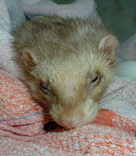
Here is our little friend nice and cozy right after her surgery, cuddled in warm blankets. She has been given a pain injection and is falling asleep.
Whenever we perform an exploratory surgery (called a laparotomy) on an animal we check other internal organs for disease. This is especially important in ferrets due to their propensity for having other diseases.

This normal spleen is being exteriorized for examination for any problems

This slightly large spleen in a ferret has a nodule that should be biopsied
We knew there was an enlarged spleen in this ferret because we could palpate it during an examination. This enlarged spleen was verified by a radiograph. Just because the spleen is enlarged in a ferret does not mean it is causing a problem. Ferrets get a spleen condition called hypersplenism, which causes a large spleen and is normal for a ferret if everything else is ok.

The black arrows outline the enlarged spleen. A kidney (K) is also visible overlying the spleen
This spleen was biopsied to determine whether this was a normal enlargement, called hypersplenism, or whether it was cause by cancer.

We have a very detailed page on spleen disease if you want to learn more.
Concurrent Disease of Ferrets with Adrenal Gland Disease
Another very important organ to check in ferrets is the pancreas due to a disease called insulinoma. In this disease the pancreas secretes too much insulin, the opposite of sugar diabetes (diabetes mellitus).
Our surgeon is carefully palpating the intestines to check for intestinal cancer, and also the pancreas (the peach colored semi-circular area in the center of the intestines) for nodules that could indicate insulinoma
 This ferret had liver cysts
Now that you know how we perform this surgery lets look at an abbreviated video of a ferret with an adrenal gland tumor and an insulinoma. You will also get a chance to see how we make a bloodless incision with the laser.
Medical Treatment of Ferret Adrenal Disease
Some ferrets are not good candidates for surgery, so we will use medical therapy on them. Also, we use this medical therapy after surgery. Medical therapy does not cure the disease, it only controls it. The effectiveness of these therapies varies from ferret to ferret.
As time and our knowledge has progressed, in many cases medical therapy is the way to go as opposed to surgery. Newer drugs that last longer after injection are giving “cure rates” that rival those of surgery, so we are using this medical therapy much more commonly.
Medical therapy does not always work, so in some cases surgery is still the way to go. This is especially true in ferrets as opposed to other animals since ferrets are prone to insulinoma and cancer, and these can oftentimes be identified during a surgery to remove an adrenal gland tumor as you saw previously.
Since it is the left gland that is more commonly involved (and much more easily removed than the right), it might be advantageous to remove the left, then put a ferret on one of these drugs if the right one starts causing a problem. These drugs are even used successfully in spayed and neutered ferrets as a prevention of the disease in the first place.
The drugs used are a synthetic GnRH analog, and is used currently to reduce testosterone in dogs as a contraceptive. When given as a injection it is slow release, and has helped reduce the typical symptoms of ferret adrenal disease
Deslorelin
This is a long acting drug that is implanted under the skin and has become our current drug of choice. The symptoms of adrenal disease in ferrets start diminishing within a few weeks, and reach maximum effect within 6 week. The injection can last anywhere from 6 months to well over one year before needing an additional injection. It is preferred over Lupron due to cost, availability, and MUMS (minor use-minor species) labeling from the FDA.
There has even been some evidence that using Deslorelin (Suprelorin) in November or December can even prevent the onset of adrenal disease. Much of this information needs to be refined as we learn more about this disease.
Lupron
Another medication used to treat this condition is called Lupron (leuprolide), originally used to treat testicular cancer in men and endometriosis in women. We also use it to treat birds like cockatiels that are excessive egg layers, and can even become egg bound.
Lupron (Leuprolide) is a GnRH analog (remember the physiology section above) that minimizes the secretion of the sex hormones from the adrenal gland. Lupron does this by suppressing the production of FSH and LH from the pituitary gland. This decreases levels of estrogen and testosterone, and symptoms diminish. It will decrease symptoms but not the size of the tumor.
It is usually given as an injection that lasts several months at least, all depending on the dose. We titrate the dose based on symptoms, age, weight, blood results, body condition score, and client compliance. Some ferrets exhibit a swelling at the injection site. This is of no cause for alarm and will usually resolve.
We have maintained ferrets on Lupron successfully for many years after the left adrenal gland is removed. Long term effects of the chronic use of this drug are unknown. In some cases we also use only medical therapy, particularly if a ferret is not a good surgical candidate due to age or other conditions that are so common in ferrets.
Adrenal tumors are so common in the United States that some veterinarians that work with large numbers of ferrets even use Lupron as a protective plan with the following criteria:
Indoor ferrets that are exposed to unnatural photoperiods- give 1 mg Lupron Depot 4-month every 6-8 months starting at 4-8 months of age.
Outdoor ferrets exposed to natural photoperiods- 1 mg Lupron Depot 4-month annual in Feb or March in North America starting at 4-8 months of age.
Viadur, a 1-year Leuprolide implant is planned for the future.
Miscellaneous drugs with potential
The following medications have been used with varying success on adrenal disease in ferrets. Much more work needs to be done before we can recommend them routinely. There is concern that long term use of them can cause damage to organs like the kidneys. No long terms studies have been performed.
Mitotane
This is one of the primary drugs used to treat dogs with Cushing’s. It is sometimes in conjunction with Lupron. It seems to show variable success only with cortical adenomas, and does not help adenocarcinomas. Its seems to work by chemically reducing the size of the affected adrenal gland. If a ferret has insulinoma at the same time, which is not uncommon, mitotane can cause hypoglycemia (low blood sugar) and a ferret can become comatose.
It is sometime used as pulse therapy, and symptoms of hair loss sometimes return after stopping Mitotane. As general rules, this drug, or in combination with Lupron, is only used on older ferrets with both adrenal glands affected and also a poor candidate for surgery due to other diseases occurring at the same time.
Melatonin
Melatonin inhibits the release of GnRH, suppressing the amount of LH and FSH released into the bloodstream. It theoretically helps regulate hormonal control in a ferrets normal circadian rhythm of light and dark. It is in the form of a long acting injection or daily oral medication.
Melatonin can be used to stimulate hair regrowth, it does not suppress the adrenal tumor. If used prior to a physical exam it can mask symptoms, and cause the adrenal gland to become even larger before surgical removal.
It can cause lethargy, and should not be used in smaller ferrets. Ferrets can develop a tolerance. It should be used as an adjunct to Lupron.
Casodex
This oral drug blocks binding sites to testosterone. It will help minimize symptoms of adrenal disease, especially straining in the male ferret. It needs to be used one week on, one week off, for life. More information is needed before we can recommend this drug.
Arimidex
This oral drugs interferes with the enzyme pathway that converts testosterone to estrogen. It should not be used with Casodex since the two drugs will negate each other.
Prognosis for Ferrets with Adrenal Disease
Most ferrets that have this surgery regain hair growth and do well for many years. Medical therapy can work well in many ferrets.
Even though most adrenal tumors are benign, recurrence can occur. A tumor can appear in the adrenal gland that is not removed at the time of surgery, and symptoms can recur.
Return to Ferret Adrenal Diseases page