
Introduction

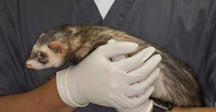
Ferrets are lots of fun, even in the exam room, as attested to by this ferret trying to climb up Dr. P’s sleeve during an exam
Almost all ferrets in the United States come from a company called Marshall Farms. They routinely spay, neuter, and descent all ferrets before distributing them to pet stores and other dealers.
In years past, before all female ferrets (called Jills) were routinely spayed, an enlarged vulva in a sick ferret was a sign of a ferret that did not mate when it was in heat. Being an induced ovulator, meaning Jills would only release eggs from their ovaries when they mated, Jills that did not mate would secrete an excess of estrogen from their ovaries for a long period of time.
This excess estrogen was toxic to the bone marrow, and could cause a severe life-threatening anemia. The technical term was aplastic anemia. The anemia would show up as severely low RBC’s (red blood cells), HGB (hemoglobin) or HCT (hematocrit),
Please confirm that your female ferret has been spayed upon purchase or however you obtain your female ferret.
If your female ferret develops an enlarged vulva it should be brought in for an exam immediately to determine if it is in heat or possibly has an adrenal gland problem.
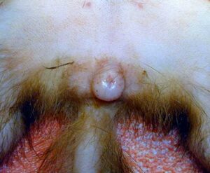
This female ferret that is laying on her back has an enlarged vulva
A female ferret can have an enlarged vulva because it is in heat (very rare since all of them are spayed prior to purchase) or because a remnant of ovary is left after the surgery.
Ferret Reproductive Physiology
Female ferrets have a unique reproductive system. Most female mammals have a heat cycle the phase in and out of, whether they mate or not. Ferrets are induced ovulators, and will stay in heat until they mate. While in heat a female ferrets secrete high levels of estrogen. If this hormone stays in the blood for a prolonged period of time, as what occurs when the female does not mate, it will affect the bone marrow.
The white blood cells are not produced in adequate numbers, and the ferret becomes much more susceptible to an infection. Also, a serious anemia will arise, and will be life threatening if not corrected. If your ferret is not being bred then it must be spayed or the problem of life threatening bone marrow suppression will present itself when it goes into heat.
We monitor for anemia on our routine blood panel by checking what is called a CBC (Complete Blood Count). The CBC checks WBC’s (White Blood Cells), RBC’s (Red Blood Cells), platelet, and even checks for blood parasites.

This ferret does not have anemia. As a matter of fact the RBC’s are high, although this is not unusual in a ferret due to splenic contraction. This is considered normal if the ferret is not ill.
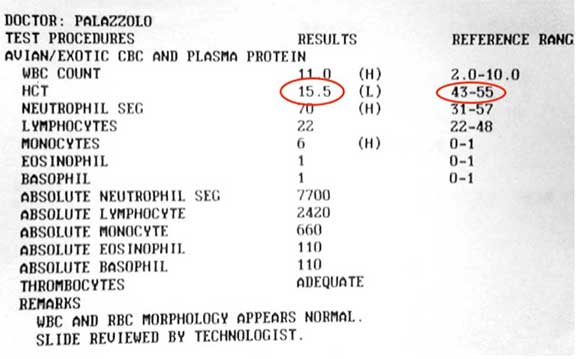
This ferret has a hematocrit (HCT) of 15.5 %, typical of a ferret with aplastic anemia. It should be at least 43%. This is severe and would require a blood transfusion.
Prevention of Anemia due to Estrogen
The best way to prevent this fatal anemia is to spay (OVH or ovariohysterectomy) your female ferret. An OVH is removal of both ovaries and uterus, and this prevents it from coming into heat (estrus) or getting pregnant.
Pre-Surgical Preparation
This preparation occurs long before surgery. We ask that you bring your ferret in for an exam and a pre-anesthetic blood panel one week prior to surgery to make sure it is healthy and ready for surgery and anesthesia. You will get to meet one of our surgeons personally and ask any questions. Before you leave a custom surgical plan will be discussed with you.
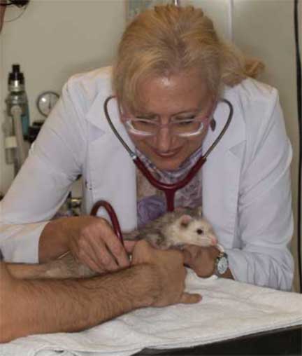
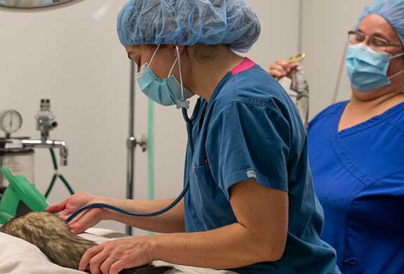
Dr. Kennedy performing a physical exam on a very wiggly patient

Part of this pre-anesthetic exam includes a blood panel
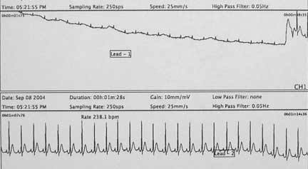
We also recommend a pre-anesthetic EKG (electrocardiogram). This one is normal.
If you would like to learn more about the fascinating world of heart diagnostics in animals follow this link and learn more about EKG’s, and even see one in action.
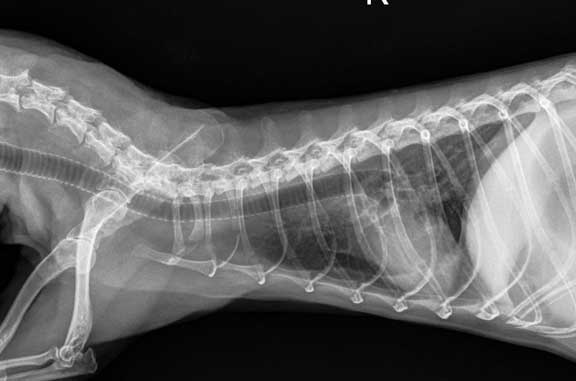
A pre-operative radiograph (X-ray) might also be taken if your doctor thinks it is indicated after the physical exam. The white structure in this chest radiograph is the heart, and the horizontal dark tube from left to right is the trachea (windpipe). This is where we put the breathing tube (endotracheal tube) when we administer oxygen and anesthesia.
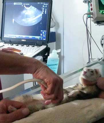
If we suspect adrenal gland disease an abdominal ultrasound will look at the adrenal glands
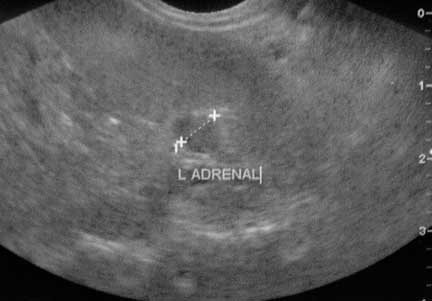
This is what an enlarged adrenal gland looks like during ultrasound
Once our surgeon is satisfied that our patient is ready for anesthesia and surgery an IV (intravenous catheter) is placed to allow us to give fluids and injectable sedatives.
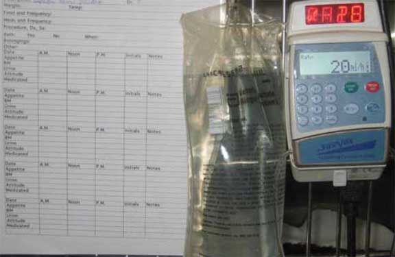
A special type of fluids are given with an IV pump for accurate dosing

Our patient’s temperature is constantly monitored. This is important in small surgical patients since they are prone to hypothermia (low body temperature).
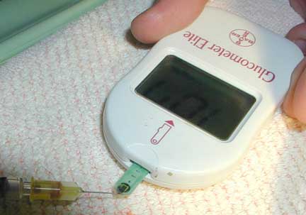
We also monitor the blood glucose levels since they are prone to hypoglycemia also
Pre-surgical Preparation
This is a sterile abdominal surgery, and our surgeon starts the pre-surgical process by using special soap to clean his hands.

We use a special surgical scrub and brush to scrub before putting on sterile gloves
While our patient is being anesthetized our surgeon is already in our surgical suite setting up instruments. Our surgeon is ready to start before our patient is at a proper plane of anesthesia. Once the anesthetist gives the green light the surgery starts immediately. We want our surgeon waiting for his patient, not the other way around. All of this is to minimize anesthetic time.

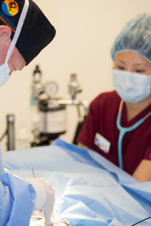
Our doctor waiting patiently prior to surgery
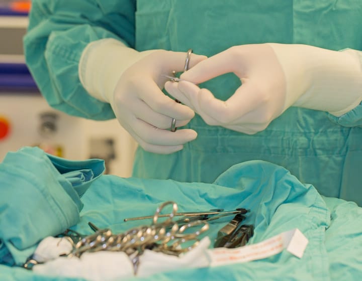
While waiting instruments are prepared so that the surgery can start as soon as the patient is brought into the surgical suite
Anesthesia
When everything is to our satisfaction we will administer a sedative. This will calm the pet down and make the administration of the actual anesthetic, along with post operative recovery, much smoother. Once a pet is anesthetized, prepared for surgery, and had its monitoring equipment hooked up and reading accurately, the surgery can begin.
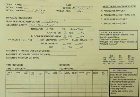
We use a detailed anesthetic form for every surgery
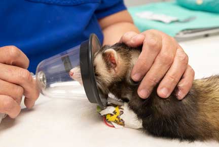
We initially administer anesthetic to our sedated pet via face mask
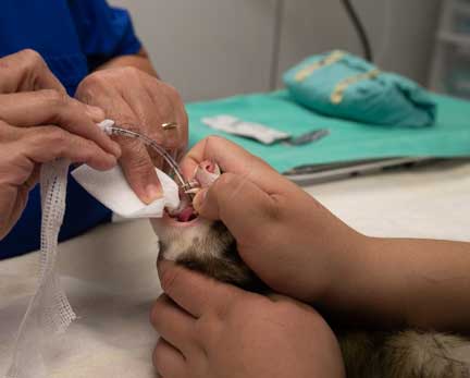
All our ferret surgery patients are then intubated for accurate administration of anesthesia and oxygen. You saw where this tube goes in the chest radiograph earlier in this page.
Our surgeon and anesthetist work closely together

We use a special stethoscope (called an esophageal stethoscope) that is passed down the esophagus and can give us a clear sound of the heart
We keep a close tab on important physiologic parameters for all of our surgeries. Monitors like this give us an early warning of an impending problem.

This machine monitors:
Temperature
Heart Rate
Heart rhythm
Oxygen saturation
Carbon dioxide level
Respiratory rate
We also carefully monitor oxygen saturation on our surgical patient with a special and small pulse oximeter
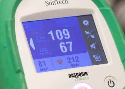
We constantly monitor blood pressure and heart rate. This ferret has a blood pressure of 109/67, and a heart rate of 212 beats per minute. This is normal for a ferret under anesthesia.
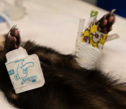
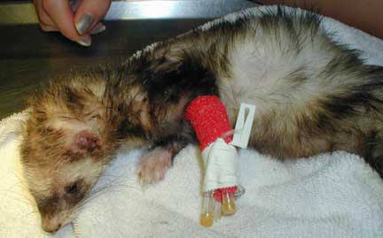
You can see the pediatric blood pressure probe on the right leg, and the IV catheter on the left leg, of this ferret under anesthesia
In addition to our monitoring equipment our anesthetist stays “hands on” in monitoring important physiologic parameters.
Our anesthetist has access to the patient at all times before, during, and after the procedure
Ferret Spay (OVH) Surgery Procedure
Our patient, wrapped in a warm towel, being brought into surgery
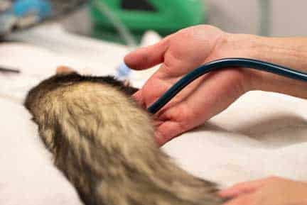
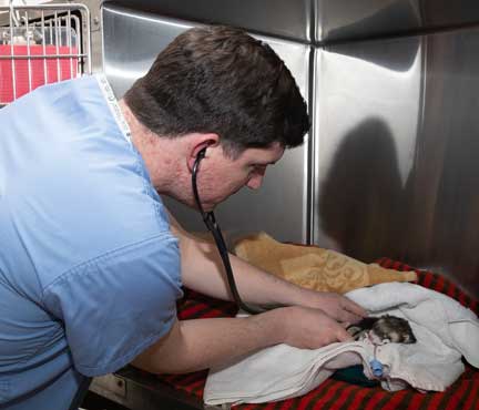
Our doctor is listening to this ferrets heart with a stethoscope just prior to starting the surgery. This is called auscultation.
Our surgeon and anesthetist work closely together
We use a special stethoscope (called an esophageal stethoscope) that is passed down the esophagus and can give us a clear sound of the heart
In this picture the skin incision has already been made and we are using a scalpel to incise the linea alba.
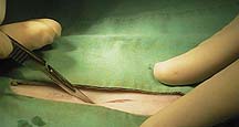
A scissors is used to extend the linea alba incision. Now we have access to the abdominal structures.
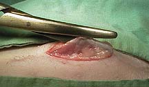
This incision gives us a full view of the abdomen and its structures. Before we can find the uterus we commonly encounter fat, intestines, spleen, and even urinary bladder.
The uterus needs to be exteriorized from the abdomen for the spay to proceed. In this view one horn of the uterus is exposed. The arrow points to the location of the ovary, buried in fat.

Sutures are placed around the ovary and it is removed form the abdominal cavity along with the rest of the uterine horn.

The same procedure is performed on the other ovary. The black arrows point to the ovaries that were just removed. The blue arrow to the right points to the location where the uterus will be removed from the body. Everything to the left of this blue arrow is removed during the procedure.

This is what remains at the cervix after it has been sutured and the rest of the uterus removed. This small amount of remaining uterus will be placed back into the abdomen.

It is very important that the linea alba is properly resutured. a hernia with actual spillage of abdominal organs can occur if the sutures aren’t placed properly.

When all of the sutures have been placed (in this case they are stainless steel) there is a solid seal in the linea alba. These sutures cause minimal tissue reaction and have tremendous holding ability. They will stay with this pet for the rest of its life, and will even show up on an x-ray of the abdomen.

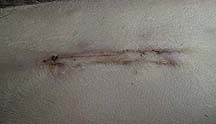
Several different types of sutures can be put in the skin incision. This type, called subcuticular, makes is difficult for the ferret to chew them out because the sutures are under the skin surface. These sutures will dissolve by themselves, so there is no need to remove them.
Post-Operative Care

Our surgery patients get therapy laser treatment to minimize pain and aid in the healing process
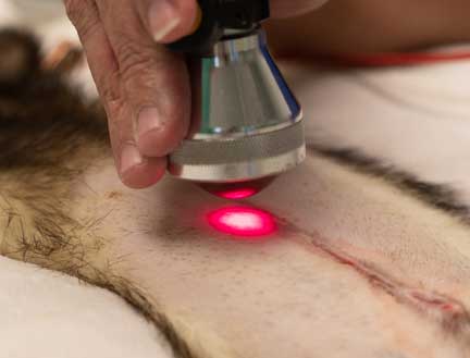
The laser penetrates the skin at the incision and works on the tissue below
At this point in the surgery a pain injection will be given and the patient allowed to wake up slowly.
After surgery it is off to recovery
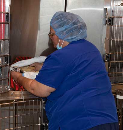
While in recovery she is monitored closely by the nursing staff
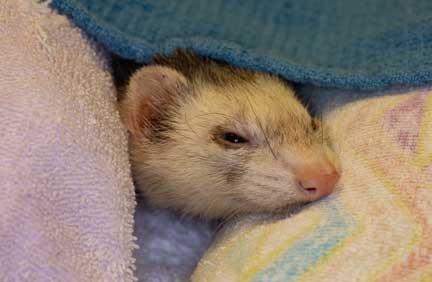
Our patient cozy and warm in her blanket, comfortably resting after surgery
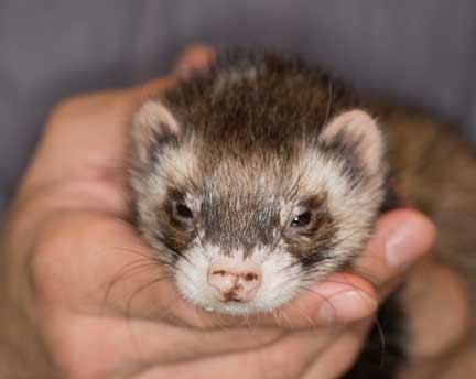
Time to go home after an eventful day
Student Externs
We are a registered teaching hospital that has trained many veterinary students over the last 35 years in our Externship Program. Getting to be a part of a ferret spay is invaluable to them since they do not get this exposure in veterinary school. The work they do with us is hands-on, and starts them on their way to getting the skills necessary to work with ferrets.
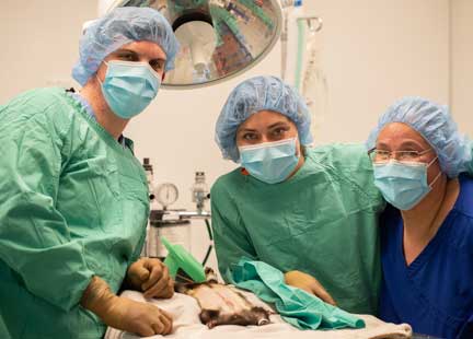
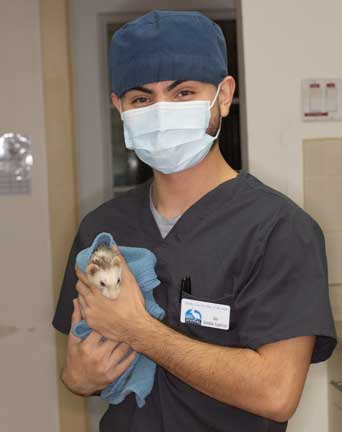
Our extern David Harris from the U. C. Davis veterinary school with our surgical team
David learning how to monitor our ferret spay patient in recovery
Our externs are required to post their daily experience at our hospital on our Facebook page. It is called the Extern Daily Diary. If you go to our Facebook page and scroll down for many years you will see the many posts they have written.
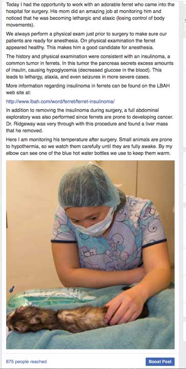
This student made her Facebook post on a ferret that had an insulinoma surgery
Ferrets make wonderful pets. They are prone to several diseases; the two most important you need to be aware of if you want to have a ferret for a pet are:
Return to Ferret Diseases Page