
Introduction
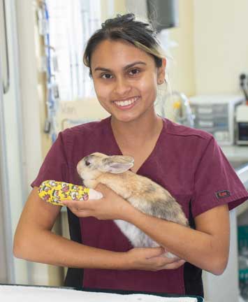
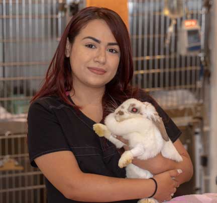
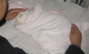
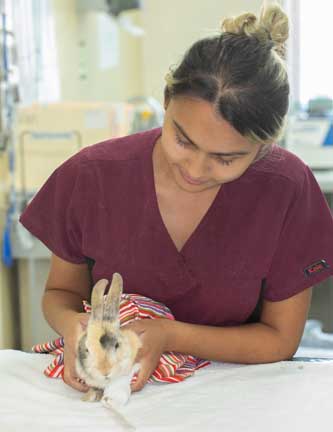
This rabbit, being held by one happy nurse, has a fractured forearm (radius and ulna) that is being cared for with a splint
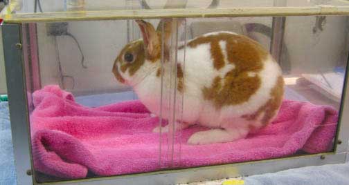
This page is about a rabbit with a much more severe fracture of the hind leg. If not repaired surgically it would debilitate this rabbit, and probably lead to eventual amputation.
Rabbits have powerful muscles in their hind legs. If they get stuck or trapped in something they can kick out and cause a bone to break. When this happens in the hindleg like the rabbit in this page, it almost always necessitates surgery.
What is a Fracture?
A fracture is a broken bone. It can be just a crack in the bone requiring only rest, a splint or a cast, or the bone can be displaced and surgery is needed with either an intramedullary (IM) pin, or a bone plate or special pins. There are more detailed assessments of bone fractures:
- Open fracture means a piece of the bone is sticking out through the skin. Some people use the older term of “compound fracture” for this.
- Comminuted fracture means the bone is in pieces. The femur in the radiograph below is an example.
This femur is in 10 pieces
- The fracture can also be spiral or transverse
Physical Examination
Rabbits do not tolerate pain well, and anyone that has broken a bone can attest to how painful a broken bone is. You can literally go into shock from the pain. We handle these rabbits with kid gloves, use pain medication immediately, and put on a temporary splint when feasible to help minimize the severe pain.
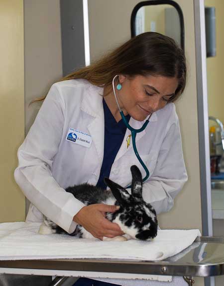
Our doctors are very skilled and experienced at rabbit medicine
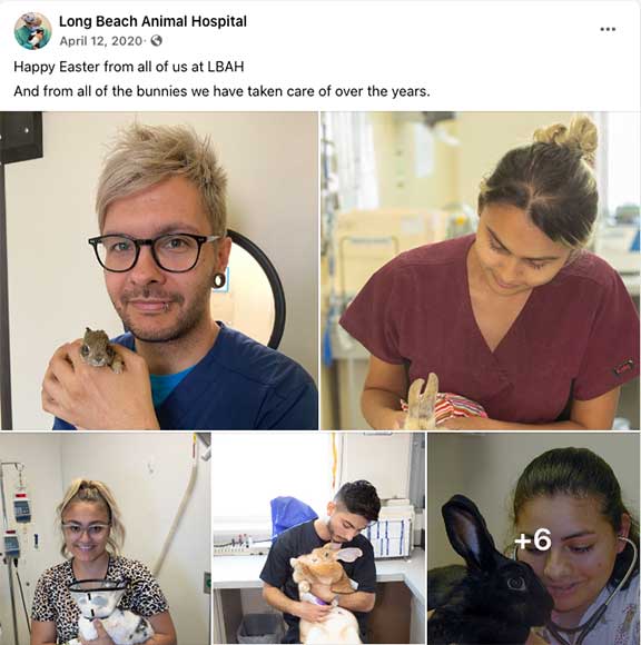
Our staff is very adept at handling rabbits based on the sheer volume of them presented to us as you can see from this Facebook post
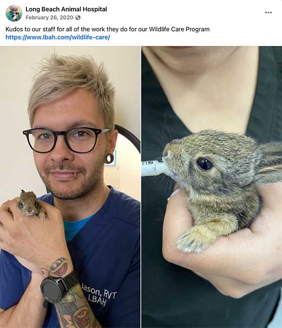
We see lots of little bun buns in our Wildlife Program where we provide free care for injured wildlife
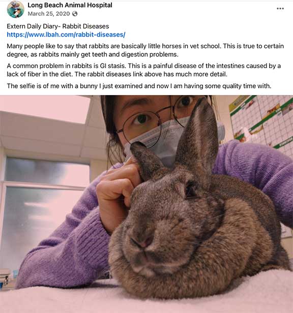
We also train our veterinary student externs in proper and gentle rabbit handling. This is a Facebook post from one of the students when she did her “Daily Diary” , reporting what she learned for the day. You can see many years of these student extern “Daily Diary” posts by clicking on our Facebook page link. There is lots there, so pace yourself!
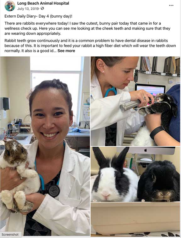
Another “Daily Diary” FB post from a student at the U. C. Davis veterinary school
Radiology
After a thorough physical exam and stabilization, radiographs were obtained. Sometimes surgery is needed, as in this case of a rabbit with a distal transverse fracture of the femur (thigh bone). In this page you will see a femur fracture surgery.
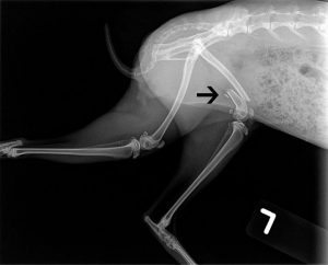
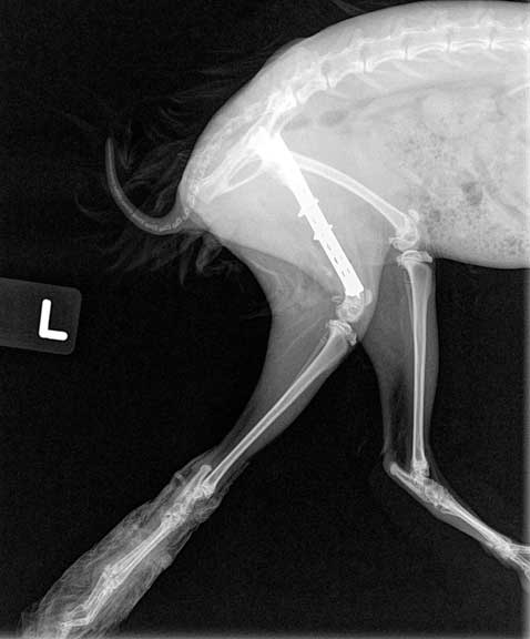
The arrow points to the fracture on this lateral view
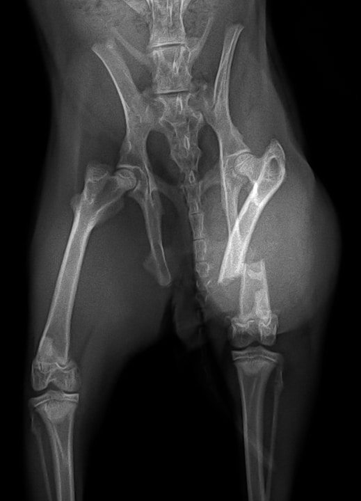
The fracture from a different view showing its severity
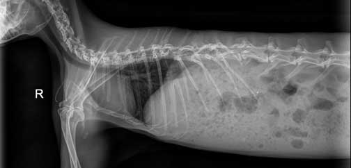
Any animal with a traumatic injury like a broken bone can have a problem elsewhere from that trauma that is not apparent. This is why we take a radiograph of the rest to the patient, especially the chest. The chest is towards the left, with the dark are being the lungs, and the white structure the heart.
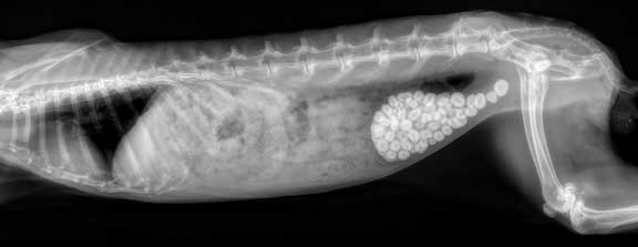
You just never know what you are going to find when you radiograph a rabbit! This one has a urinary bladder filled with stones! Click here to learn more about bladder stones in other species.
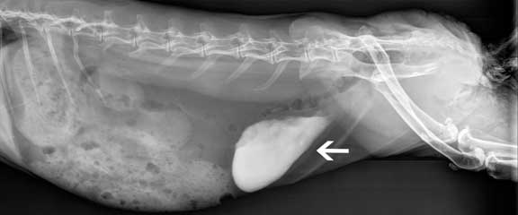
This one has a problem with calcium sludge in the urinary bladder. Our Calciuria page has more details.
Pre-Surgical Preparation
Blood Panel

A normal blood panel on a 6 month old rabbit
Surgical Preparation
If the surgery day physical exam is normal we will perform the surgery. Our surgeon starts the pre-surgical process by using special soap to clean his hands.
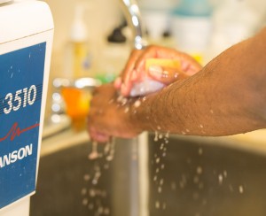
He washes his hands several times with the surgical soap and brush before putting on sterile gloves
While our patient is being anesthetized our surgeon is already in our surgical suite setting up instruments. Our surgeon is ready to start before our patient is at a proper plane of anesthesia. Once the anesthetist gives the green light the surgery starts immediately. We want our surgeon waiting for his patient, not the other way around. All of this is to minimize anesthetic time.

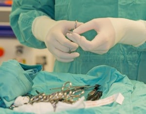
After scrubbing, gowning, and gloving, our surgeon opens up the surgical pack
He then confirms the instruments are sterile with a sterilometer. This is an indicator strip inside the surgery pack confirming the instruments have been sterilized properly.
While our patient is being anesthetized our surgeon prepares the instruments

Dr. Ridgeway sorting instruments while waiting for his patient to come in to surgery
This is a sterile surgery, and this concept is particularly important in orthopedic surgery since an implant (bone plate or IM pin) is inserted for months at a time, or even indefinitely in some cases. Also, bones heal slowly, and an infection can fester during this long healing process. If an infection gets started in a bone it tends to become deep seated and hard to control.
In orthopedic surgery we use specialized instruments. The primary ones are wires, bones plates, and IM pins:
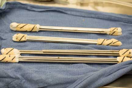
Sterile IM pins ready to be implanted
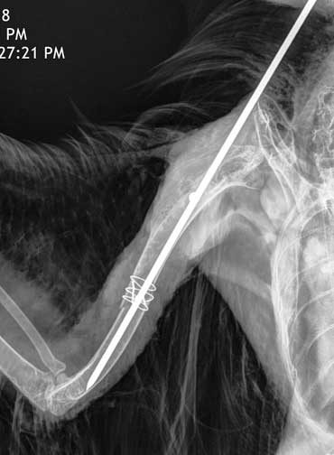
What the pin looks like in a hawk from our Wildlife Program with a fractured humerus. The wire that is giving additional support is called a cerclage wire. It also needs to be sterile, since it stays in this bird forever. The pin was taken out a few months later. You can see the whole story of this hawk, from beginning to end, including the surgery and release.
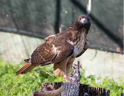
To give you a preview of this story, here he is at the rehabber convalescing
What a bone plate looks like on a dog’s fractured femur. Lots of screws are used, and they all need to be sterile since the plate and screws might stay in a long time.
Anesthesia
Before any of our rabbit patients are anesthetized we give them plenty of TLC to minimize stress
Anesthetic monitoring is important is such a small animal, and especially in rabbits that have such a small lung capacity compared to other animals of comparable size.

The heart and lungs (within the red circle) are tiny compared to the size of the abdomen. This ratio between lungs and abdomen is much smaller than in other animals, and needs to be closely watched during anesthesia.
Before surgery we carefully examine our rabbit patients to make sure they are ready for anesthesia, and the pre-operative pain medication is working
When the rabbit’s pre-anesthetic blood panel and physical exam are completed, it is anesthetized and brought into surgery.
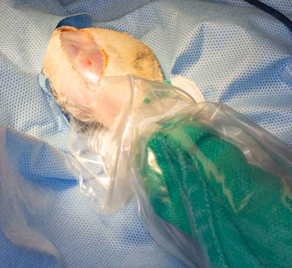
We use a special gas anesthetic that is gentle and safe. This is the induction chamber that is filled with 100% oxygen prior to administering any anesthesia. We do this to make the anesthetic safer.
Rabbits need to be monitored carefully during anesthesia. Because of this we constantly monitor their temperature during and after the surgery.
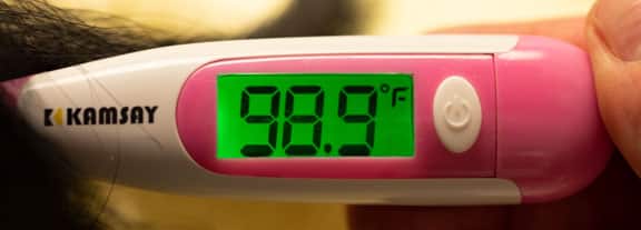
We use digital thermometers for their accuracy and speed

Our surgical patients are kept warm with a circulation warm water blanket and additional warm fluids

We also use a hot air blanket to keep our patients warm when needed

Detailed records are kept of the anesthesia and surgery
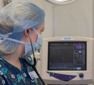
We use a sophisticated anesthetic monitor to constantly assess important parameters

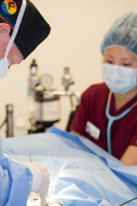
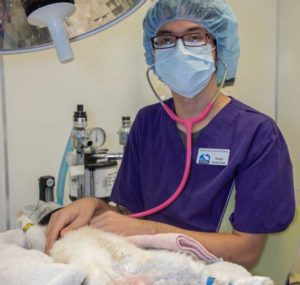
We have a team of people that are present for our rabbit surgeries

Our anesthetist works closely with our surgeon to make sure just enough anesthetic is given at the lowest possible dose
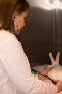
Our anesthetist is using a stethoscope to monitor the heart. We use this hands-on approach in addition to all the monitoring equipment.
We have a detailed page on anesthesia for much more information on how we anesthetize a wide variety of species.
Surgical Process
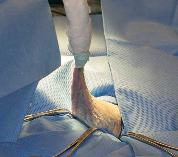
The technique of taping the leg up is the standard in how we clip and clean the leg prior to surgery
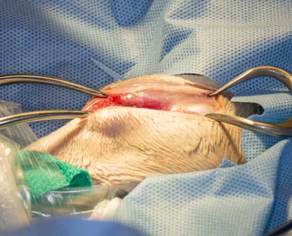
Our surgeon palpates the fracture through the skin to find the best place to make the initial incision. You can see the foot double wrapped in a special towel, and also plastic wrap at the lower right of the photo. The foot is not a part of the surgery, and draped this way so there is no contamination.
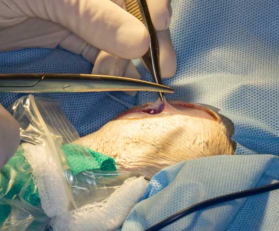
The initial skin incision exposes the muscle layer below. There is minimal fat under the rabbits skin so this initial skin incision has to be done carefully or else the scalpel will cut into the muscle.
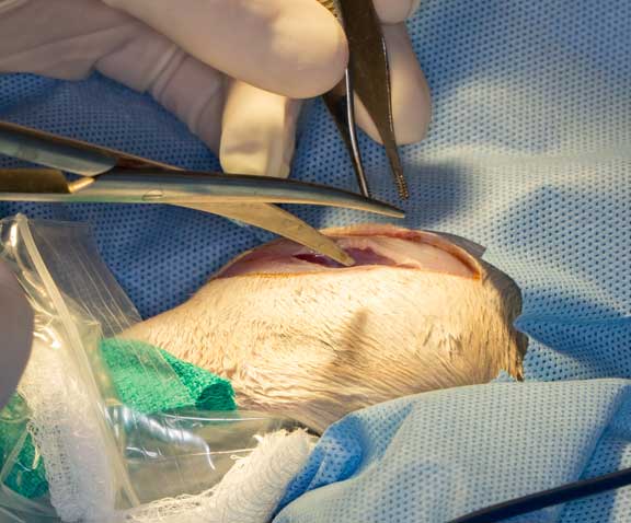
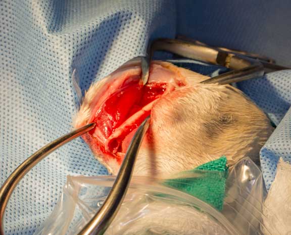
A layer of tissue over the muscle is cut with scissors
After carefully dissecting through specific muscle planes a special instrument is used to spread the tissue for better access to the fracture.
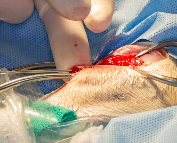
With the bone exposed our surgeon now assess the damage. Even though the radiographs taken before surgery give us substantial information, decisions on how to repair the fracture are only decided at this point. You can see the tip of the fracture at the lower left.
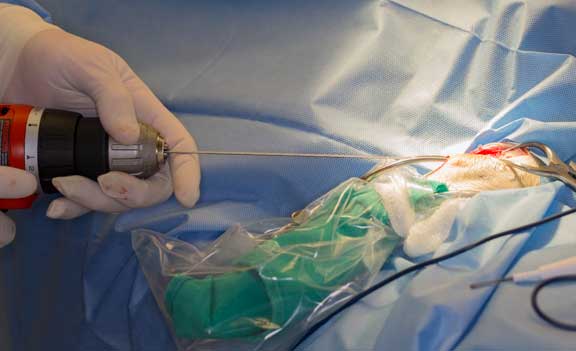
A stainless steel intramedullary (IM) pin is placed down the shaft of the bone. You can see the pin entering the open end of the bone on the left. This is the first part of stability of the fracture site, and is guide for the alignment of the 2 fractured ends.
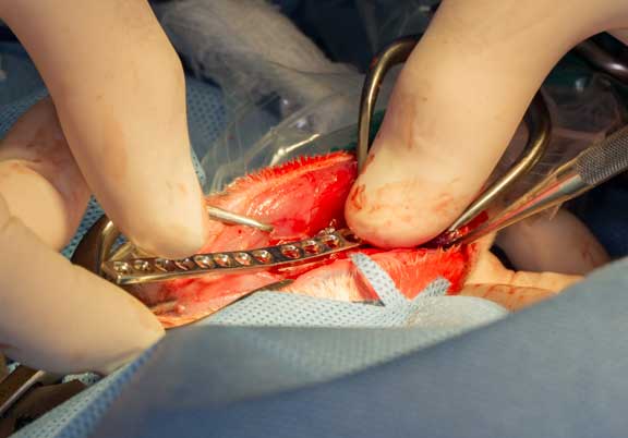
Once the pin is in place a stainless steel bone plate is hand molded to the contour of the femur
A hole is drilled into the bone through one of the holes at the end of the plate
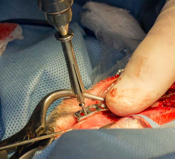
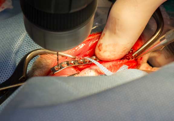
A tap-less screw is then inserted manually so the surgeon can give the right amount of pressure to make it snug without cracking the bone
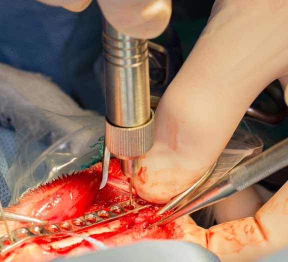
This is repeated at the other end of the bone plate. You can see our surgeon measuring how deep a hole has been drilled into the bone. This helps pick a screw that is just the right length.
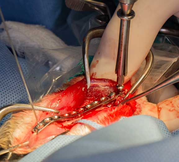
The correct size screw is now placed. This is the 2nd aspect of stability of the fracture site.
After putting in 3 screws our surgeon decided that more screws might damage the bone. Two cerclage wires are now used, which is the 3rd aspect of stability. When our surgeon is happy with the stability from this wire, he cuts it.
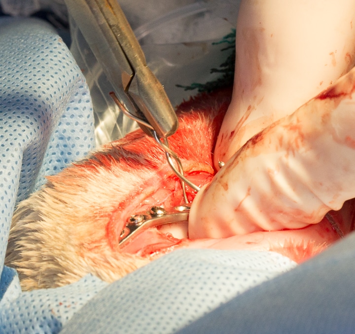
Starting the first cerclage wire
Tightening it to just the right tension
Testing it prior to cutting it short
Cutting the extra length with a special stainless steel cutter
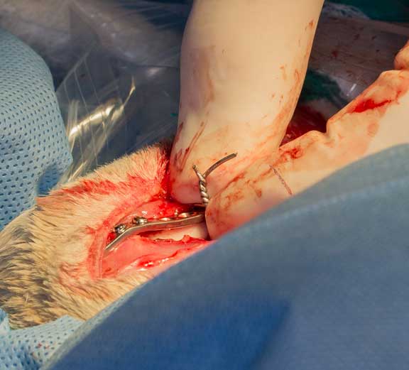
Putting in the second cerclage wire over the plate
Tightening it just like the first one
What this second cerclage wire looks like prior to cutting it shorter
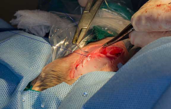
Suturing the tendinous tissue over the muscle that was initially incised to get access to the fracture site
Suturing of this layer complete
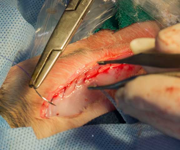
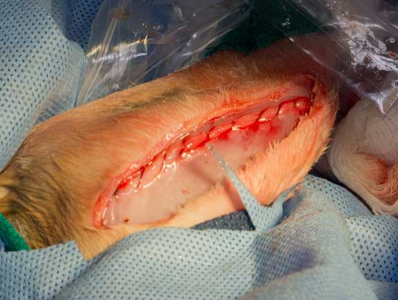
Preparing to suture the skin
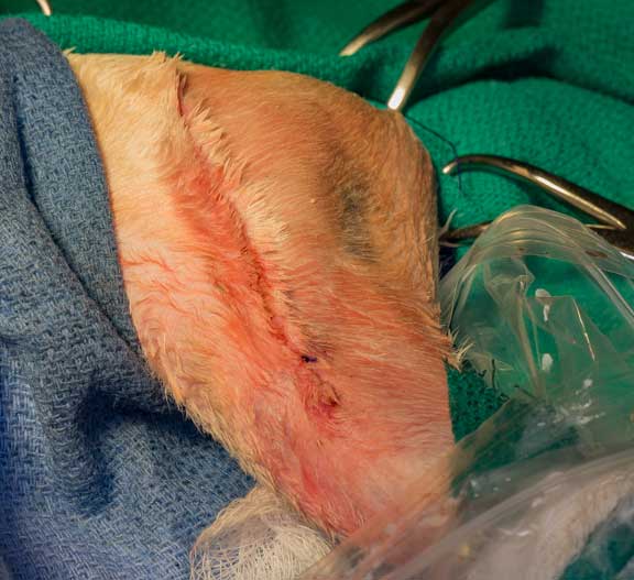
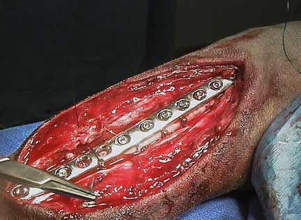
The final appearance of the incision
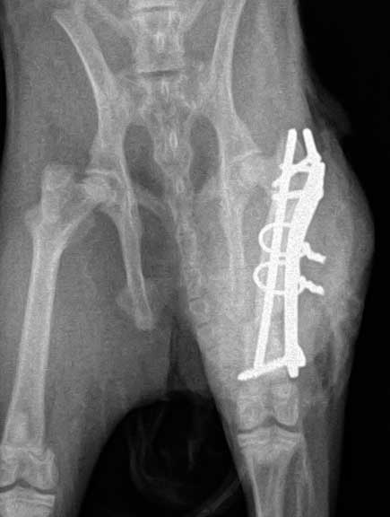
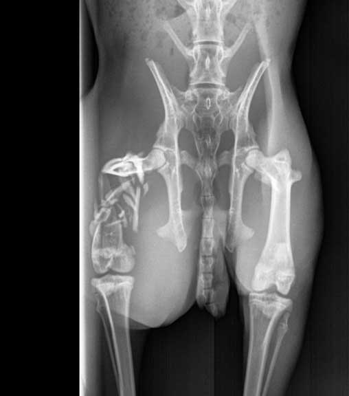
Can you visualize the 3 screws and 2 cerclage wires over the bone plate in this post operative VD (venture-dorsal) radiograph of the pelvis?
This is the lateral view of the plate
Post-Operative Care
Before our patient wakes up it is given a pain injection and give companion laser therapy. It was also given a line block, which is an injection of long acting local anesthetic called marcaine (bupivicaine) so our bunny wakes up pain free. This line block lasts 8 hours, and hits the pain before it even starts, a huge advantage in pain control.
Before fully awake we use our Companion Laser to stimulate the healing process and decrease post operative swelling at the incision. This also aids in pain control at the incision site.
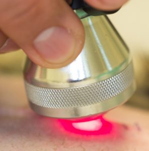
This special laser aids in healing, and decreases pain and inflammation after the surgery
It must be kept in mind that although the bone has screws in it, the stability of the femur now that the fracture ends are not rubbing each other and the muscle, greatly aids in pain control.
Our patient is closely monitored in the immediate post operative period
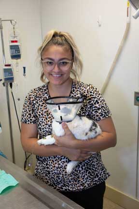
Rabbits tend to be chewers, so it is common for us to put on an E-Collar (aka the cone of shame) as soon as they wake up
After surgery our rabbit patients are wrapped in a towel and closely watched by our staff until fully awake
We have a page called Home Care of the Surgical Patient to give you some pointers once you get your patient home.
Prevention of Rabbit Leg Injuries
Many rabbit leg and spine injuries occur when a rabbit is startled and jumps off of a table or out of someone’s arms. Always have a towel on a table so the rabbit feels stable and secure and is not inclined to kick out its back legs and break its spine or jump off the table. The same thing holds true when you hold your rabbit. It needs to feel comfortable and secure in your arms, or else it will kick out and potentially fall to the ground breaking a bone or its spine.
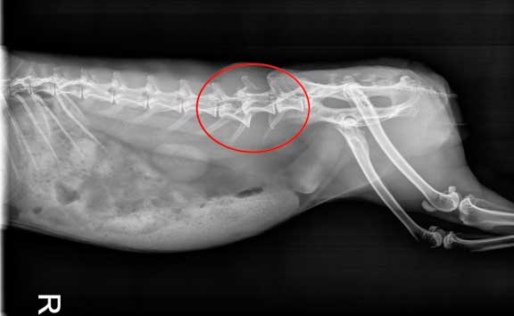
A fractured lumbar vertebrae like this can lead to paralysis of the rear legs
If you are not sure how to hold or restrain your rabbit our staff will give you a demonstration of the proper technique.
Wellness Care
Rabbits are wonderful pets that are not long-lived relative to dogs and cats and people. Also they can hide symptoms of disease naturally as part of their normal instincts to evade detection by a predator. This stresses the importance of bringing your rabbit in for a yearly Wellness Exam so that we can look for early warning signs of disease and run a blood panel to check the internal organs.