
The most common reptile brought to our hospital is the green iguana. The primary reason owners bring in their ill green iguana is because it has developed a disease called metabolic bone disease (MBD), also known as nutritional secondary hyperparathyroidism (NSHP), or just secondary hyperparathyroidism (SHP). This page shows how we diagnose and treat Iguana Bone Disease (NSHP, MBD).

This Iggie decided he just wanted to hang out and drink our free coffee

This big boy shows how much bigger they can get. Unfortunately, some of these male iguanas are this large because they were fed a diet too high in protein when they were young, which will affect their kidneys later in life.
You can learn more about our standard of care of sick reptiles from the Association of Reptile and Amphibian Veterinarians.
The main cause of this disease is poor husbandry, especially an inadequate diet. Unfortunately, the literature abounds with erroneous information on the precise needs of these creatures. This outdated and incorrect information continues to be propagated by individuals and organizations with good intentions but limited knowledge.
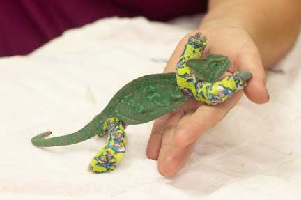
Iguanas are not the only reptile that encounters this problem. We find it in many types of chameleons, lizards, turtles, and tortoises, although not as often as we do in iggies.
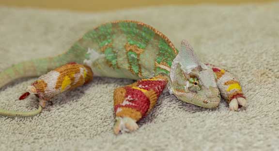
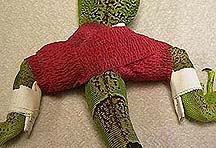
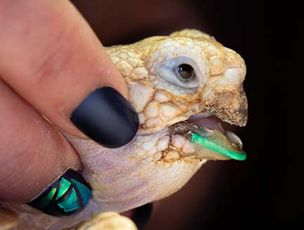
Click on this chameleon link and learn how this cute little guy, with many broken bones that we splinted, has MBD.
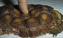
This disease also occurs in tortoises. The shell is soft due to inadequate nutrition in this baby California Desert Tortoise.
It does not occur in carnivorous reptiles like snakes and monitor lizards very often because of their lower requirement for UVB light, and the whole prey they consume provides a proper diet. In essence, they are carnivores, while the other reptiles are vegetarians.
Cause
This disease has many factors that work together in causing this condition. The primary reason iguanas develop this disease is due to a diet too low in calcium. More specifically, the ratio of calcium to phosphorous (usually the phosphorous is too high) in their diets is inadequate to promote growth and sustain normal physiological functions.
The high phosphorus from the inadequate diet drives down the calcium level in the bloodstream. As a result, they become very ill which you will learn about soon, and can even succumb to the disease.

Low calcium is called hypocalcemia. High phosphorus is called hyperphosphatemia.
In older reptiles this disease manifests itself as renal secondary hyperparathyroidism (RSHP) due to compromise of the kidneys. The symptoms are the same as NSHP. In this case the blood sample shows high calcium and sometimes phosphorus.
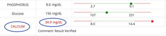
In this blood panel the calcium is high, like this one that is 34.9 (up to 14.4 is normal). It is so high that the lab double checked the number, which is why it says “Results Verified”. When we multiply the calcium x phosphorus we get a ratio that tells us about the kidneys. This ratio is high due to kidney failure.
Other factors that exacerbate the poor diet problem are common in most households that have iguanas. Inadequate exposure to direct sunlight (not through glass), not keeping the humidity at 90% and not keeping the temperature at 90 degrees F all add to the problem:
Sunlight of a specific ultraviolet frequency is needed to produce vitamin D3 by the iguana’s skin. This vitamin is needed for the absorption and utilization of calcium in the diet. No matter how much calcium there is in the diet, without this vitamin the calcium would not be be absorbed or utilized . This is why milk that we drink is fortified with vitamin D. Black Lights and other artificial ultraviolet lights are helpful, but they can not replace sunshine.
In order to maintain normal bodily functions (ability to digest food, fight infections, etc.) an iguana needs to maintain a high body temperature. Since they are reptiles, they maintain this temperature by absorbing the heat from their environment. They cannot produce enough internal body heat like birds and mammals can when placed in a cold environment. Also, the precursor to vitamin D needs to be at the proper temperature to be converted to the active form of the vitamin.
In the semi arid environment in some parts of the country (southern California for example), or the heat needed to warm homes in the winter, many iguanas live in a perpetual state of dehydration. This dramatically interferes with their physiology, and predisposes them to many problems.

This is a typical Iguana cage that is inadequate. There is no branch with leaves to bask on, no access to any sun, let alone through the glass, and inadequate humidity. The bowl of water does not give enough moisture, and the heat lamp that is present in the corner does not supply proper ambient heat. Putting dirt in the bottom of the cage is a good idea.
They type of caging and lighting needs to take into account the unique physiological needs of each reptile species. For example, the UVB light source for lizards needs to be above the cage because only then can it stimulate their pineal gland, which regulates their daytime basking.

This is the pineal gland on the top and middle of the head of this iguana that is looking towards the left
Caging and husbandry also needs to take into account the feeding behavior of each reptile species. Some need the food in trees and vegetation, while others need food on the ground. Water bowels need to be at different sizes and depths to accommodate individual preferences.
All of this means that you need to fully understand the husbandry of the reptile species you are contemplating on having as pet if you want it to live a healthy life. Diseases like MBD are serious, life threatening, and lead to significant suffering that is not necessary, and ignorance of the needs of each individual reptile and amphibian is no excuse.
Symptoms
Iguanas with this disease have many problems. Initially they might be sluggish, unable to life their trunks very high off the ground, and not able to walk well.
The bones might be swollen or soft. The jaw might be swollen (called lumpy jaw by some people) because nature is trying to bring in supporting tissue to make up for the lack of strength to the bones of the jaw. The same thing happens to the bones of the legs, and when the problem is severe enough, or has gone on long enough, the bones of the arms and legs can fracture (called a pathologic fracture) all by themselves. You saw that in the chameleon picture above.
Some of these iguanas will be unable to walk properly due to spinal cord damage, and many of them will be more susceptible to common infections because they are too weak to develop a proper immune response. As the bones of the jaw become weaker it becomes impossible to eat, further exacerbating the problem. In severe cases the blood calcium level becomes so low that tremors occur.
They might have distended abdomens and bones leading their owners to the erroneous conclusion that their pet is fat and sassy, and receiving an adequate diet. Growing iguanas and females laying eggs have a greater need for calcium and might be more prone to this problem.

A proper diet, with the proper Calcium:Phosphorous ratio, is needed for healthy eggs

Females with eggs might not have the strength to lay them. This picture is from a surgery to remove toxic eggs in an egg bound iguana.

Lumpy jaw occurs when the body brings fibrous tissue to the area to stabilize the weak bones. A lump at the jaw can also be caused by an abscess.

The fold of skin on the side of this iguana is evidence of dehydration
This Iguana has such a low calcium level that the muscles are twitching. This is called tetany, and is a serious sign. The two most common causes of tetany in an Iguana are MBD and Kidney failure.
This Iggie is so weak it cannot move properly
Diagnosis
A diagnosis of NSHP is made based on history, physical exam findings, and radiography. The history might indicate a diet of iceberg lettuce, dog or cat food, or packaged iguana meal. Swelling of the jaw and legs, low body weight, weakness, dehydration, hypothermia, poor appetite, and lethargy all might be noted on physical exam.
X-rays (radiographs) are very helpful in the diagnosis. We assess all organs, especially the bones.
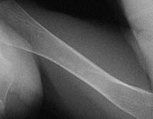
This is the femur (thigh) bone of a healthy iguana. Compare it to the diseased one below.
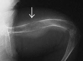
This is the thinning (arrow) that occurs in this disease. In addition to poor bone strength, this iguana has an infection.
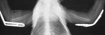
This iguana has a severe form of the disease. The spine is deformed which has interfered with the nervous system, so it is unable to walk or eat well. This creature is gravely ill. To let a creature deteriorate to this point is a crime.
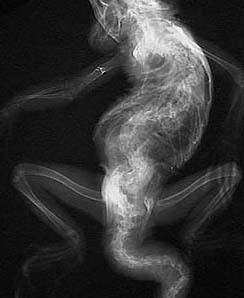
An x-ray reveals the extent of the curvature problem to the spine
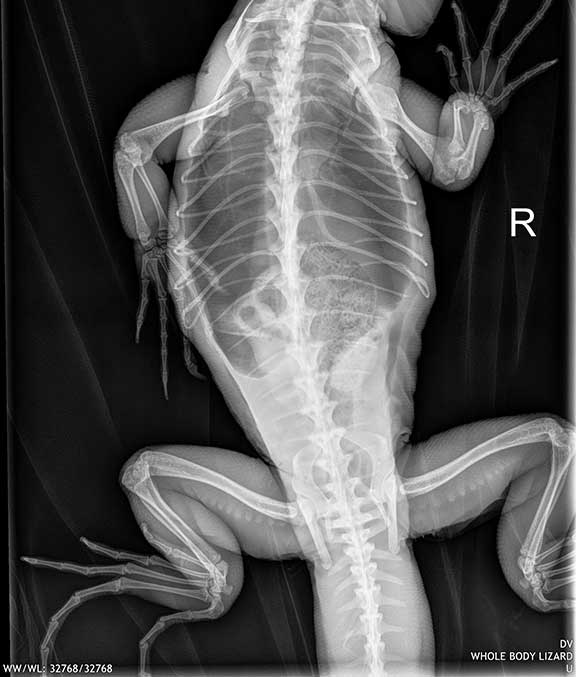
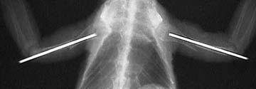
This is the radiograph of a normal iguana. Notice how straight the spine is and how strong the leg bones are.
Even though this is a problem of low calcium level, blood samples show normal calcium levels commonly. This is because the body is absorbing calcium from the bones.
Kidney failure in iguanas can cause RSHP as explained earlier. When there is kidney failure, the blood panel might show the problem.

We had an indication this sick iguana had a kidney problem when we palpated the large kidneys in the abdomen (it’s actually called the coelomic cavity in reptiles). The kidneys are far back in the coelomic cavity compared to most other animals.

A radiograph show enlarged and calcified kidneys

The calcium and phosphorous are both very high, commonly seen in kidney failure. A picture of an iguana with a calcium even higher than this was show earlier in this page.
Getting blood on a reptile all depends on the species and their size. Their veins are not visualized or palpated anywhere near as readily compared to dog and cats. You need to go by “feel” sometimes.

On iggie’s we take it from the underside of the tail where there is a venus plexus

On tortoises we take it from the jugular vein
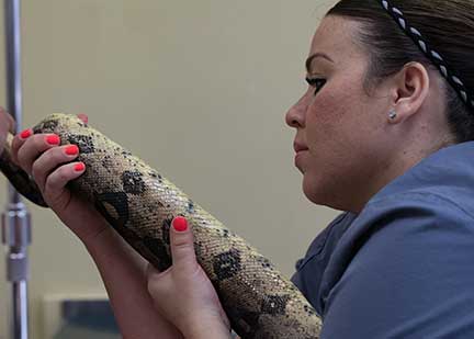
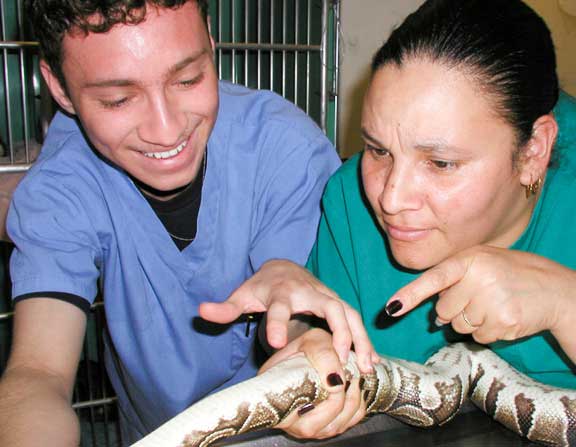
On snakes we take it directly from the heart. The real challenge is finding the heart!

It is obvious in this picture- you do see it, don’t you?

If you need help we can pull out the ultrasound to find it

Good job, you found it!
Our nurses sometimes have a contest to see who can find it first. Loser buys donuts for everyone!
Treatment
Iguanas that are diagnosed with NSHP are usually very ill and often need to be hospitalized in a special warm and quiet room. During hospitalization they are given fluids to correct dehydration, a special liquid diet, injections of vitamin D3, injections of calcium, oral calcium, and antibiotics if they have an infection.
After they are stabilized in the hospital they are sent home with calcium supplements, antibiotics if needed, and their dietary deficiency is corrected. They need to return weekly for at least several weeks for vitamin D3 injections and calcitonin injections.
Those that have pathologic fractures are splinted. A typical splint applied when both rear legs have pathological fractures
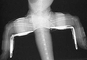
An x-ray of this splint shows the padded paper clips that are used for support
This different case was referred to us. The splints on these front legs are inadequate, as evidenced by the displacement of the fractured ends.
We performed surgery in order to correct this problem. These pins will be removed in 1-2 months.

This iggie shows how the femur bones look after a successful treatment to stabilize fractures. The excess bone around the cortex of each of these femurs is called callus.
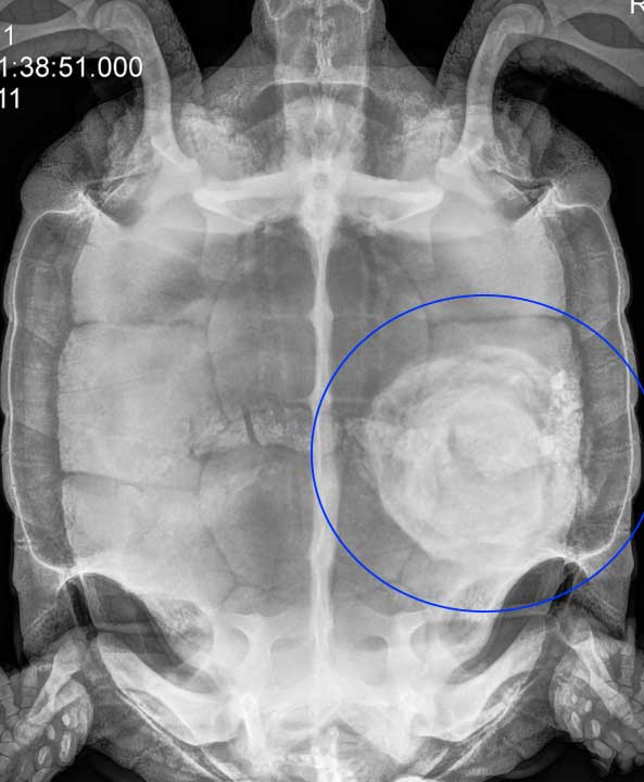
We sometimes see severe MBD in other species besides iguanas. Look at this California Desert Tortoise (CDT) radiograph and you will spot the problems at the arrows. Can you tell what that white thing is at the jaw?
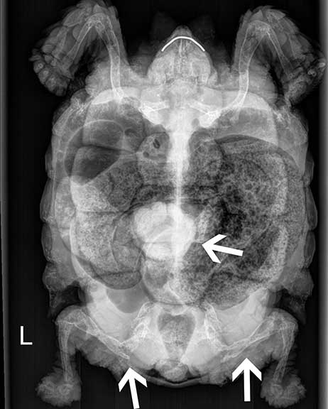
The lower arrows point to severely diseased and fractured rear legs (femurs) of this CDT. This upper arrow points to an impaction in the intestines, although a bladder stone can look like this also.
The jaw was also fractured, and in the radiograph above the while line is the McGyver type of splint we fashioned for it.
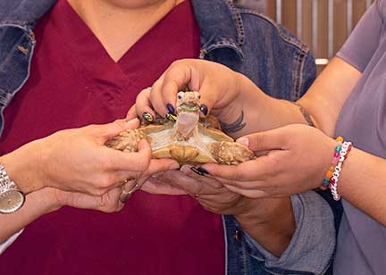
It was a three person job to brace the jaw and prevent it from spontaneously fracturing (called a pathologic fracture)

Front view of the jaw splint
This is also a serious problem in chameleons as you saw at the beginning of the page. Click on the picture above to learn a whole lot more, and watch videos of their eyes moving independently!
Prevention
It must be fully understood that iguanas are ectothermic animals. This means they are highly dependent on their environment for their normal physiological functions, much more so than birds and mammals. They come from Central America where the temperature and humidity are consistent- 90% humidity and 90 degrees F. In addition, they bask in the direct sun many hours each day. If these conditions cannot be replicated, then these animals should not be kept in captivity. Ignorance of their needs is no excuse.
Their diet should consist heavily of dark green leafy vegetables in order to have the proper ratio of calcium to phosphorous. Contrary to what you may read elsewhere, these animals are not omnivores, they are strictly vegetarians, even when they are young. Therefore, high protein diets (dog food, cat food, protein supplements) are not to be fed to them. These high protein diets will cause them to grow bigger and faster, but will also cause their kidneys to fail. Vitamin supplements that contain calcium should be given weekly.
The following list describes would should and should not be fed. A combination of several items from the “Should Be Fed List” need to be fed, not just one or two items. For small iguana’s it is helpful to dice up your vegetables into very small pieces so that they don’t eat only a few items.
Foods That Should Be Fed
|
Foods That Should Not Be Fed
|
Prognosis
This disease is correctable and preventable. If a pet iguana is brought to us in an advanced state of the disease then the prognosis is not good. Otherwise, we are able to return a large percent of them to relative normalcy if our full treatment regimen is followed. After the immediate problem is corrected it is mandatory to provide the optimum environment for their proper quality of life.
We have a very interesting page on how we remove a bladder stone from a California Desert Tortoise. You get to watch the surgery, and even see the beating heart during the procedure. Click here and enjoy!
It is not uncommon to see stones this large!
Return to Reptile Diseases Page