
This page describing kidney disease is very thorough, with pictures and information on anatomy, physiology, and pathophysiology, in addition to symptoms, diagnosis and treatment. If you pace yourself and set aside the time to read it you will get through it. We have a summary page on kidney disease if this page is too detailed.
Even though this page is thorough, the physiology of the kidneys is complicated, and only a summary of how the kidneys work is presented on this page. How the kidneys regulate water consumption and urine production are controlled by complex and highly refined interactions between plasma osmolality, fluid volume in the bloodstream, the thirst center in the brain, the kidneys, the pituitary gland, and the hypothalamus. It is part of what is called the RAAS- Renin-Aldosterone-Angiotensin-System. You will learn about a small part of this system in the physiology system explanation to follow.
One of the more common conditions encountered in pets, especially as they age, is kidney (renal) disease. It is estimated that anywhere from 7%-20% of dogs will get this problem sometime in their lives. This disease is more prevalent in cats, and 30% of them over 10 years of age will get chronic kidney disease (CKD). It is also known as chronic renal failure (CRF) or chronic interstitial nephritis (CIN).
Once a pet has CKD the changes in the kidney are irreversible, so it is important to catch this disease early to mitigate its progression. Early intervention with diet is to the key to keeping this chronic problem from progressing more rapidly than it should.
There are early warning signs that should be monitored closely once your dog reaches 8 years of age:
Any degree of decrease in appetite- this is a crucial aspect of this disease and needs to be watched closely.
Weight loss- you should weigh your pet weekly to catch this early since it is easily missed.
Excessive drinking and urinating- Monitor how much water you put in the water bowl to determine a change here
Risk factors predisposing your pet to getting kidney disease:
Dental disease
Episode of cystitis (bladder infection of FLUTD)
Neutered male
Recent bout of dehydration
We have information on treatment towards the end of this page if you want to bypass all the background information and skip right to it.
IRIS Classification System
We use the IRIS (International Renal Interest Society) system of classification at the Long Beach Animal Hospital to better diagnose and treat CKD. IRIS is a group of veterinary kidney specialists throughout the world that have studied this disease extensively, and have set standards for diagnosis and treatment.
The IRIS system stages kidney disease from Stage I to Stage 4. At the end of this page there is a link to this organization for more information on how they stage kidney disease.
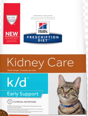
As the problem progresses to IRIS stage I kidney disease, k/d Early Support is used.
It also comes in different sized cans and bags
As we monitor changes in the kidneys you will learn about in this page, we will change the diet to regular K/D©. This food is the gold standard for kidney disease in dogs and cats, and we have used it for over 40 years.

There are many versions of this food
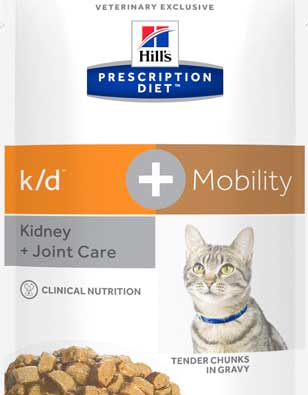
If your elderly cat also has arthritis, which is common in this species, there is a mobility version of k/d to help
These foods work, and it is important to use them to slow down the progression of CKD so your pet leads a long and healthy life. Please do not let someone at a pet store, or a neighbor, give you advice on how to feed your pet with kidney disease. They do not have the knowledge needed, some of which you will read about on this page, or the experience treating pets with this problem like we have for many thousands of patients.
In addition, they give their advice freely without your pet’s body condition score, its blood panel and urinalysis, and its X-ray and ultrasound results. This is inappropriate, and they should not be giving any advice, as well intentioned as it is.
These Hill’s foods have an increasing progression of less protein. They still contain the important amino acids, called essential, that are needed by the body to make proteins. In the dog and cat the essential amino acids are:
- Methionine
- Arginine
- Threonine
- Tryptophan
- Histidine
- Isoleucine
- Lysine
- Leucine
- Valine
- Phenylalanine
- Taurine (cat only)
These essential amino acids are obtained only from the diet. The other amino acids, which are produced by the liver, and not needed in the diet, and are called non-essential.
The point of these foods is to decrease the total protein in the diet by restricting the amount on non-essential amino acids. By having less of these non-essential amino acids, the kidney have less work to do to deaminate the protein. Less work for the kidneys to do is always a good thing, since the kidney has lots to do already, that you will learn about in this page.
Our Nutrition Page has general information on pet foods for more detail.
25% of the blood ejected from the heart on every heartbeat goes directly to the kidneys, a testimonial to how important the kidneys are to health. When a pet has chronic kidney disease there are many issues that need to be addressed for a successful outcome. You will learn about them in more detail later in this page. The more important ones are:
- Protein and phosphorous regulation
- XS protein in the urine
- Elevated creatinine in the bloodstream
- High blood pressure
- Anemia
- Dehydration and electrolyte imbalance
- Low pH in the bloodstream
- Stomach and intestinal ulcers
- Low body condition score- your pet is underweight
In the IRIS staging system we are closely monitoring several important parameters:
- Creatinine trends
- Protein in the urine (this is one reason why a urinalysis is important when we run a blood panel on your pet)
- Blood Pressure
- Body condition score
- SDMA (symmetric dimethlyarginine) trends

This pet has a normal BUN and creatine, so initially the kidneys seem OK on the blood panel
The urinalysis shows a protein level of 1+ on this same pet
Even though the kidneys are normal on the blood panel, the protein of 1+ is an early warning sign that the kidneys might not be functioning at 100%. Just changing to a food like Hills c/d or k/d Early Support can make a big difference on a pet like this if started early in the course of the disease, and before other problems related to CKD appear. This emphasizes the importance of Wellness Exams for dogs starting at 8 years of age.
In addition to these parameters, the following general parameters are also checked for early signs of chronic kidney disease. These parameters are also used to monitor progression and success of treatment:
- Small kidneys on abdominal palpation
- Radiographic or ultrasound evidence of small kidneys
- Decreased urine specific gravity
Pets that have kidney disease commonly have other problems that need careful attention if the kidney problem is to be treated successfully. Some of these other common problems are:
- Hyperthyroidism
- Heart disease
- Inflammatory Bowel Disease
- Dental disease
- Sugar diabetes (diabetes mellitus)
- High blood pressure (hypertension) – leading to blindness.
On occasion a dog with acute kidney disease can become so dehydrated and hypothermic (low body temperature) that they can go into shock. This is a medical emergency requiring immediate veterinary care.
The Long Beach Animal Hospital, staffed with emergency vets, is available until the evenings 7 days per week to help if your pet is having any problems, especially shock, pain, breathing hard, or bleeding. Always call us first (562-434-9966) before coming in. This way our veterinarians can advise you on what to do at home and so that our staff and doctor can prepare for your arrival. To learn more please read our Emergency Services page.
Medical Terminology
Several medical terms are used when describing kidney disease:
azotemia– excess nitrogenous waste products in the bloodstream
hypokalemia– low blood potassium level
hyperkalemia– high blood potassium level
anemia– low red blood cells
BUN– blood urea nitrogen
GFR– glomerular filtration rate
hypertension– high blood pressure
hypophosphatemia– low blood phosphorous
hyperphosphatemia– high blood phosphorous
polydipsia– excess drinking
polyuria– excess urinating
PU/PD– polyuria and polydipsia
CRF-chronic renal failure
ARF– acute renal failure
Renal anatomy
The kidneys are such a vital organ that 25% of the blood that enters the circulatory system from each heartbeat goes directly to the kidneys through the renal artery. With such a high metabolic rate the proper functioning of this organ is critical to health. The high metabolic rate and importance of this organ makes the kidneys susceptible to many problems.
The kidneys are located in a specific area of the abdomen called the retroperitoneum. This area is a small indentation at the top of the abdomen just underneath the spinal vertebrae. This location, in addition to being surrounded by a fat, affords added protection to this vital organ.

The kidney is normally covered in a layer of fat
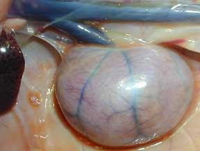
This is a kidney with surrounding fat removed. You can see the dark liver on the far left, and the renal vein as it leaves the kidney and merges with the vena cava (the dark blue horizontal structure at the top)
The blood from the vena cava flows into the liver and then directly to the heart. This is the blood that has been filtered of impurities and is re-entering the circulation. You can not see the renal artery or ureter, they are buried in the white fat at the top of the kidney.
Blood enters the renal artery and flows into the nephron where it is filtered. The blood from the renal artery that has been filtered now flows out of the renal vein where it goes back into the circulatory system. The impurities that the nephron filters out of the blood collect in the renal pelvis and eventually out into the ureter in the form of urine.
The primary functional unit of the kidney is the nephron. Each kidney has upwards of one million nephrons, so obviously they are microscopic in size. Every nephron is a self contained unit that can form urine by itself. Not all nephrons are used at the same time, which gives the kidneys the capacity to increase their workload if called upon.
This reserve capacity is lost when chronic renal failure occurs. These pets outwardly appear normal, but have greatly reduced ability to adapt to changing physiologic needs. Being chased by a dog, not having enough water to drink, etc., can send them into a crisis, requiring immediate medical care.
This is a kidney, turned partially sideways to the right compared to the one above, as we view it with the ultrasound. The yellow line is measuring its length.
The important anatomical components of the nephron are described below:
Afferent Arteriole
This small artery is one of the many small branches that come off the renal artery as it enters the kidney. It supplies the glomerulus with blood. Eventually filtered blood returns to the renal vein.
Glomerulus
This is a collection of many small blood vessels at the end of the afferent arteriole. Normal pressure of the blood in the glomerulus causes fluid to flow into a collecting area called Bowman’s capsule.
Bowman’s Capsule
Fluid that collects in Bowman’s Capsule eventually flows into the tubules. It is in these tubules that waste products and excess electrolytes are filtered out of the fluid, and normal blood constituents like protein and glucose are absorbed back into the bloodstream. When a diuretic like Lasix is given it acts on these structures.
Collecting Ducts
At the end of the tubules is the collecting duct, where the urine produced starts to flow out of the nephron. Other nephrons deposit urine in collecting ducts as these ducts flow into the renal pelvis. From the pelvis the urine flows into the ureter and bladder.
Renal Physiology
The kidneys have a profound affect on almost all the physiologic processes of the body. The mechanisms by which the kidneys perform these functions is extremely complex, and only the more important ones will be discussed:
Fluid Regulation
In relation to the kidneys, the brain monitors bloodstream levels of water, waste products, electrolytes, and red blood cells. The circulatory system also has receptors like the brain to monitor blood volume also. If the water level is too low, as occurs with dehydration, the brain secretes more of a hormone, called ADH (anti-diuretic hormone), into the bloodstream.
As a result, the kidneys excrete less water into the urinary tract, retaining more fluid in the bloodstream to counteract the dehydration. The brain also increases thirst simultaneously. The end result is less urination. The urine that does get excreted is more yellow than usual due to a greater concentration of waste products being excreted in relation to the amount of water being excreted. The only thing we notice is that we urinate less and it is more yellow in color.
As we drink water to quench our thirst and rehydrate, the body notes this change and the brain secretes less of the hormone called ADH. Now when we urinate more water is excreted by the kidneys, and our urination occurs with a dilute urine in greater quantity.
So, the ability to concentrate the urine and dilute the urine is an important function of the kidneys. It is a fine tuned mechanism that is closely regulated to maintain optimum amounts of fluid in the bloodstream and organs.
As a fun fact, it is the inhibition of ADH by alcohol’s depression effects on the brain that causes excess urination when drinking alcoholic beverages. Eventually this excess urination causes dehydration, leading to that inevitable curse called a hangover.
The kidneys also secrete a hormone called renin. Through a complicated set of biochemical pathways this ultimately leads to an increase in salt (sodium) in the bloodstream. Sodium pulls water towards it, so more sodium means more fluid in the bloodstream. It will have an effect on blood pressure, which you will learn about later.
All of this is a part of the RAAS (Renin-Aldosterone-Angiotensin-System) mentioned in the beginning of this page.
Waste Product Regulation
The brain also monitors waste products that build up in the bloodstream. These waste products are the end product of normal metabolic processes, especially the metabolism of proteins. They are called nitrogenous waste products, and are measured by a blood parameter called blood urea nitrogen (BUN).
Another waste product that is closely regulated by the brain and kidneys is called creatinine. It is the end product of the metabolism of muscle. Creatinine is the most important waste product we monitor on a blood panel to give us an idea of the health of the kidneys.
The kidneys also excrete toxins and foreign substances that are introduced into the body. Almost every medication given, either orally or by injection, is eliminated to some degree by the kidneys.
The rate at which fluid flows into the glomerulus is important. This is called the glomerular filtration rate (GFR), and is measured in ml/minute. Too small a flow and waste products are not eliminated, a problem encountered during dehydration. Too much flow and normal blood constituents like protein are excreted when they shouldn’t be.
Electrolyte Regulation
Electrolytes are also of importance in relation to the kidneys. Sodium is of extreme importance in the normal functioning of all cells. It allows nerve impulses to occur and is critical in the regulation of water levels in the bloodstream. Through the release of a hormone called angiotensin the kidneys regulate fluids levels of sodium in the bloodstream.
This has a major affect on the blood pressure. Potassium is also a critical electrolyte. Potassium levels need to be kept at a very narrow range to prevent serious consequences like heart irregularities.
Hormone Regulation
The kidneys also regulate calcium and phosphorous by hormones called calcitrol and parathyroid hormone, and by regulating vitamin D. Vitamin D allows the absorption of calcium from the intestines. If the kidney disease progresses long enough the excess secretion of parathyroid hormone causes the bones to become swollen and fibrous as the body attempts to maintain a normal calcium level. This is called renal osteodystrophy.
As the bones become more fibrous the marrow is not able to produce red blood cells as effectively. This leads to weak and thin bones, as evidenced by a swollen face and jaw as the bones of the lower jaw weaken. It can occur in other bones also.
This is similar to what occurs in reptiles when they get bone disease. You can see a picture of the swollen jaw of an Iguana with bone disease. Don’t forget to come back here because we are only just getting going.
Acid-base Regulation
The pH of the bloodstream, which is a measure of acidity, is another important area of kidney physiology. The kidney regulates this acidity by excreting excessive hydrogen ions and the selective secretion and reabsorption of bicarbonate.
Red Blood Cell Production
The kidneys secrete a hormone called erythropoietin into the bloodstream. This hormone circulates to the bone marrow and stimulates it to produce red blood cells. A lack of adequate levels of this hormone will cause anemia. Toxic waste products that build up in the bloodstream decrease the life span of a typical red blood cell, further exacerbating the anemia.
And, as you already learned above in hormone regulation, the fibrous bones have less bone marrow. There can even be clotting problems due to a low number of platelets.
Pathophysiology of Chronic Renal Failure
Over the course of days, weeks, or months, normal nephrons get replaced with scar tissue, and become nonfunctional. In chronic kidney disease (CKD) this scar tissue is the result of excess phosphorous. When this scar tissue occurs to approximately 75% of the nephrons the kidneys no longer have the ability to respond to the needs of the body.
There is no longer any reserve, and all of the remaining nephrons are working full time. These remaining nephrons swell (called hypertrophy) to adapt to this increased workload. This allows them to adapt and selectively excrete or reabsorb important nutrients.
Eventually these remaining nephrons cannot keep up, and it leads to a buildup of nitrogenous waste products (called azotemia) in the bloodstream. The body compensates by increasing thirst, which causes PU/PD, and the waste products get flushed out of the bloodstream and into the urine.
Unfortunately, flushing out the waste products in bloodstream with excess thirst also flushes out important electrolytes and protein into the urine. This causes weight loss and weakness as the kidneys continue to deteriorate. The excess urination that occurs as the body tries to rid itself of these excess waste products can also cause dehydration.
Oral ulcers occur when bacteria in the mouth convert the extra uremic waste products to ammonia. Waste products that buildup in the bloodstream also have an effect on the bacteria in the mouth and exacerbate gingival and periodontal disease.
The waste products also change the pH of the bloodstream and cause ulcers in the stomach and intestines. This causes vomiting (emesis), loss of appetite (anorexia) and weight loss due to pain. This pain makes them miserable, and adds to the already significant problems they have. We want to treat before it gets to this point.
Ulcers can also be found in the mouth and tongue due to the uremia. When they occur in the stomach or intestines we cannot tell unless we do an ultrasound.
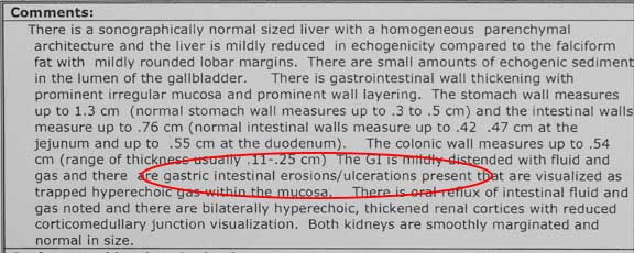
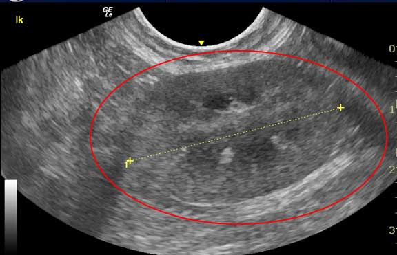
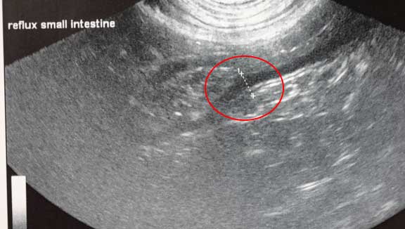
This is the ultrasound report we get when a pet has an ulcer in the stomach or small intestines
The ulcer in the small intestines
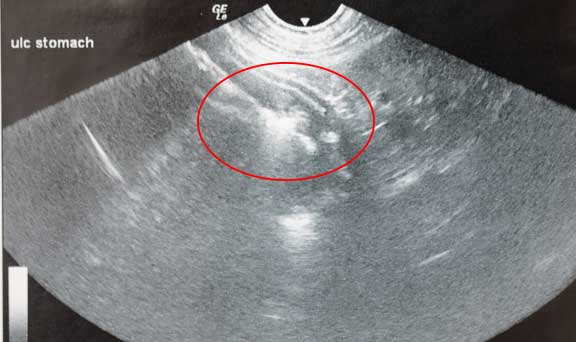
The ulcer in the stomach
Hormones are affected and phosphorous builds up in the bloodstream further adding to a pet’s woes. Eventually calcium is deposited in abnormal places, and can lead to problems with many skeletal and internal organ problems. Due to sodium imbalance, hypertension (high blood pressure) can develop. Hypertension occurs in a high percentage of animals with kidney disease.
In addition to dogs and cats, we work on a wide variety of species like pocket pets, birds, and reptiles. They also get kidney disease, although due to dramatic differences in anatomy and physiology, especially in the reptiles and birds, it is diagnosed differently. The following radiograph shows one way we diagnose kidney disease in an iguana.
This is a calcified kidney in an iguana with renal failure. Notice how far back in the abdomen (technically it is called the coelomic cavity in reptiles and birds) the kidneys reside. They are almost in the pelvis.
As the kidneys continue to deteriorate erythropoietin is not secreted in adequate quantity and anemia results. This anemia also makes a pet weak and adds to the anorexia that is usually present.The nervous system is affected by all of these problems. If the uremia is severe enough hypothermia and seizures can result.
Classification
Acute Renal Failure (ARF)
This is a serious form of kidney disease that commonly leads to death. The kidneys have an abrupt decrease in the GFR due to a toxin or loss of adequate blood supply (called ischemia). Many different disease processes can cause ARF, including anesthesia for any surgical procedure. That is why we give intravenous fluids (IV) to almost every surgical case.
Chronic Renal Failure (CRF)
This is the most common form of kidney disease we encounter, particularly in older pets. It tends to develop more slowly than ARF, so the body has time to institute corrective factors (called homeostasis) to compensate for the problem. Unfortunately, these corrective factors tend to hide early symptoms of disease. Consequently, treatment is not initiated as soon as it might be.
Again, as in many diseases we encounter, this drives home the fact that pets over 8 years of age should have annual physical exams along with blood and urine samples. This is all part of our Wellness Care.
Pets in CRF that have lost their ability to compensate for their failing kidneys can be presented to us in an acute phase, similar to ARF. These pets need hospitalization because they are ill and can succumb to their problem. Once we get them over the acute phase we treat them as CRF for the long term.
Cause
The are a multitude of causes to kidney disease. Some of these cause ARF, while others cause CRF. In some cases, ARF can progress to CRF.
Toxins
Many drugs that are used on a day to day basis can be toxic to the kidneys:
- snake and bee venom
- antifreeze
- pesticides
- herbicides
- solvents
- heavy metals
- cancer chemotherapeutic agents
- aspirin
- NSAID’s- Metacam, Advil, Ibuprofen
- anesthetics
- anti parasite drugs
- antibiotics
- blood pressure medication
The outcome of exposure to these toxins depends on a pet’s age, other disease processes that might be present, any medication your pet is currently taking, how long there has been an exposure and at what dose, along with the specific toxin. In some cases they are treated with supportive care like intravenous (IV) fluids. Other cases are treated with specific antidotes.
Some toxins, notably antifreeze ( 95% ethylene glycol) are catastrophic to the kidneys. Antifreeze is sweet tasting and is readily licked by dogs if it spills on the ground when car antifreeze is changed. Ethylene glycol is converted in the liver and kidney to a toxic metabolite that changes the pH of the bloodstream and destroys the kidneys by depositing calcium oxalate crystals in the renal tubules.
It is a medical emergency and requires specific and immediate measures if the kidneys are to be saved. Unfortunately, unless a pet owner actually observes their pet licking antifreeze, they don’t bring their pet in for care until it is very ill. In this situation the prognosis is grave, and death is common. If treated within a few hours of ingestion the prognosis for recovery is much better.
Fortunately, the antifreeze manufacturers have added a bitter taste and we do not see this disease anywhere near as commonly.
Antifreeze toxicity has several distinct phases:
Stage I
This occurs during the first 12 hours after ingestion. Pets will vomit, drink and urinate excessively (PU/PD), and appear intoxicated. It is at this stage that observant owners might bring their pet in for an exam.
Stage II
This stage occurs 12-24 hours after ingestion. Symptoms are vague and pets appear to recover. Unfortunately, this recovery is short-lived in many cases, and the problem progresses to Stage III.
Stage III
This stage appears 24-72 hours after ingestion. Pets in this stage are severely depressed, are not eating, are vomiting, and are not producing urine. When this stage appears death is imminent.
Treatment needs to be given early in the disease to be effective. Inducing vomiting and flushing the stomach out can be very helpful if performed within 1-2 hours of ingestion of antifreeze.
Intravenous fluids and diuretics are also given to maintain normal kidney function by keeping an adequate GFR. Sodium bicarbonate is given to maintain a proper pH of the bloodstream.
Antidotes are given and can be highly effective if given early enough. We give them ethyl alcohol (vodka) intravenously, and literally make them drunk. The vodka prevents the liver from converting the ethylene glycol to the toxic metabolites that destroy the renal tubules. This treatment is used in dogs also.
A better antidote, that works in dogs only, is called Antizol. It is an expensive medication, but it can literally save your dogs life.
A dog in ARF from antifreeze ingestion is a medical emergency requiring immediate veterinary care. They can collapse and go into life-threatening shock.
If you have an emergency that can be taken care of by us at the Animal Emergency Hospital Long Beach always call us first (562-434-9966) before coming. This way our veterinarians can advise you on what to do at home and so that our staff and doctor can prepare for your arrival. To learn more please read our Emergency Services page.
Cancer
Cancer of the kidneys can occur even at a young age, although it is usually diagnosed in older pets. Sometimes it arises from the kidneys (primary), much more often the cancer has spread to the kidneys from a different organ (secondary or metastatic). When primary cancer does occur it is often malignant. Fortunately, primary renal tumors are rare. Cancer of the kidneys occurs more in cats than in dogs. Click here to see a case study of how we diagnosed and treated kidney cancer in a dog.
Primary
Lymphosarcoma– This is the most common renal tumor in the cat, and those with renal lymphoma are commonly positive for the FeLV.
Adenocarcinoma- The next most common renal tumor in the cat
Transitional cell carcinoma
Nephroblastoma
Adenoma
Fibroma
Secondary
Osteosarcoma
Melanoma
Poor Perfusion
Poor perfusion means inadequate flow of blood through the kidneys, which decreases the GFR. This lack of blood flow prevents the kidneys from eliminating waste products and toxins buildup in the bloodstream. This lack of perfusion is the main pathology leading to ARF.
Heart Disease – If the heart is weak it cannot pump enough blood to the kidneys to keep them properly perfused.
Drugs – Some medications can cause constriction of the artery to each kidney with a resulting lack of perfusion
Dehydration – Inadequate fluid in the circulatory system will cause poor perfusion. Dehydration is the most common cause of poor perfusion.
Cysts
They can put pressure on normal kidney tissue and compromise the filtering ability of the nephron. These tend to be found in older male cats. When there are no symptoms they are sometimes found accidentally when checking for other problems. This is called an incidental finding. When symptoms are present, they can be mild and treated easily by drainage, or there might be compromise with the normal filtering ability of the kidneys.
A specific form of cyst is called Polycystic Kidney Disease (PKD). This tend to be a cat disease and not a dog disease. The best way to make this diagnosis is with ultrasound. Ultrasound should be used on the offspring of adult cats with PKD and before any symptoms appear.
Immune System Diseases
Bacteria, viruses, cancer, and diseases of internal organs can all set off a reaction where the immune system can interfere with the ability of the kidneys to filter properly. This is sometimes called glomerulonephritis. Symptoms range from mild early in the disease to all the signs associated with kidney failure.
A common method of diagnosis is excess protein in the urine (proteinuria) and a lack of protein in the bloodstream (hypoalbuminemia).We use ultrasound at our hospital to help in this diagnosis.
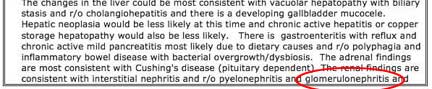
Ultrasound report of glomerulonephritis
What it looks like on ultrasound
Treatment depends on the exact cause. It might include anti-immune system drugs, aspirin, dietary change, medication to decrease blood pressure, salt reduction, IV fluids, and diuretics.
Parasites
There are 3 main parasites that invade the urinary tract and affect the kidneys:
Capillaria plica
They are threadlike worms that affect the kidneys, bladder, and urethra. Eggs of this worm that are passed in the urine are eaten by earthworms, which are then eaten by dogs to complete the cycle. In some dogs there are no symptoms, while in others there might be blood in the urine (hematuria), difficult urinating (dysuria), or urinating small amounts (pollakiuria).
This parasite is diagnosed by finding the egg in a urine sample. In most cases the disease goes away by itself within 4 months, although it can be treated. Prevention of recurring cases relies upon removal of surfaces that could harbor earthworms.
Capillaria feliscati
This is an uncommon parasite in our area that invades the urinary bladder of cats. Usually there are no symptoms, and the disease routinely resolves by itself within 4 months.
Dioctophyma renale
This parasite resides in the kidney or abdomen near the kidneys, although they have been found in the urinary bladder, urethra, ovary, uterus, and pericardium. It causes a gradual deterioration of the kidneys.
Eggs from this parasite are passed through the urine and eaten by aquatic annelids. Dogs get this parasite from eating raw fish and frogs that have eaten the aquatic annelids.
Sometimes there are no symptoms until there has been significant kidney destruction. They are diagnosed by finding the egg of the parasite in abdominal fluid or in the urine. Treatment involves surgical removal of the worms from the kidneys or abdomen.
They are difficult to control because the eggs can live in the environment for a long time. Dogs should be prevented from eating frogs and raw fish. It is possible for humans to get this disease from eating raw fish or frogs also.
Bacteria
They can ascend from the lower urinary tract and cause dysfunction in the kidneys. Leptospirosis is a specific bacteria that affects the kidneys, seen almost exclusively in dogs. Dogs get it by direct contact with infected urine through their mucous membranes. It also affects the liver.
In severe cases a dog can go into shock and rapidly die from Leptospirosis. In some cases they are sick with a fever, depression, vomiting, diarrhea, and poor appetite. There might also be muscle pain, eye problems, and respiratory problems. Most cases are chronic and might not show many symptoms.
There is a vaccine to prevent this disease which is a routine part of our DHLPP vaccine. The vaccine is highly effective in preventing this disease.
Bacteria can also cause pyelonephritis, an infection of the renal pelvis. The following bacteria are implicated:
E. coli
Staph. aureus
Proteus mirabalis
Strep. spp.
Klebsiella pneumonia
Psuedomonas aeruginosa
Enterobacter spp.
These bacteria usually ascend from the lower urinary tract. Occasionally they enter the kidney from the bloodstream. Their presence can cause constriction of the blood supply to the kidneys and destroy normal kidney tissue when attacked by the immune system. They can eventually lead to kidney failure.
It is important to culture the urine for bacteria in any pet that is diagnosed with CKD because of the damage these bacteria can do to the urinary tract.
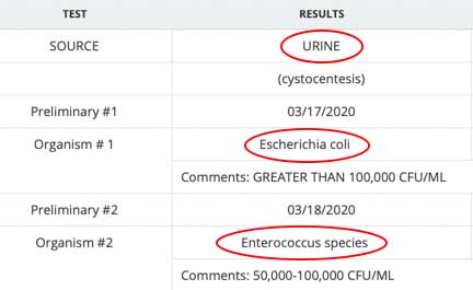
This urine revealed two different bacteria
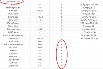
Many antibiotics were tested on the E. Coli to see if they will kill the bacteria. As you can see, the E. coli was resistant to many of these antibiotics. This is not good news.
These bacteria can cause ARF or CRF. Symptoms include fever, depression, lack of appetite, pain, PU/PD, and weight loss. In the chronic version sometimes there are no symptoms at all. They are treated with antibiotics for a minimum of 4 weeks, along with supportive care.
Amyloid
This is the deposition of fibrous protein cells in the glomerulus that interfere with the kidneys’ ability to filter. Amyloid causes the kidneys to become small and irregular. Pets with amyloidosis have typical symptoms of kidney disease.
Most dogs are middle aged or older, and it is seen in Shar Pei dogs. It is diagnosed by proteinuria, just like the immune system diseases that affect the kidney. Amyloid can be deposited slowly allowing a long life, or it can occur rapidly leading to early death. There is no specific treatment except routine supportive care of the kidneys.
Trauma
One of the more common causes of kidney trauma is when a pet is hit by a car. These injuries can be serious and easily lead to death. Radiography is helpful in making this diagnosis, although special x-rays or ultrasound might be needed to know for sure.
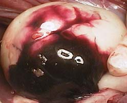
This is a bruised kidney from a pet that was attacked by a dog. The bruise covers over 1/3 of the kidney.
Symptoms of Kidney Disease
The symptoms that occur depend mainly on how long the problem has been present and the specific reason the kidney failed in the first place. Some of the more common ones you might notice at home are:
Excess urinating and drinking
This is known as polyuria/polydipsia (abbreviated PU/PD). It is by far the most consistent symptom of kidney disease. You will note this by your pet hanging by the water bowl more, and more urine in the litter pan, or your dog needing to go outside to the bathroom more often.
PU/PD also occurs in sugar diabetes and hyperthyroidism to name a few, so the diagnostic process needs to be followed to make an accurate diagnosis of a pet with symptoms of PU/PD.
In ARF there might not be any urination (called anuria) at all. This is an extreme emergency. Two of the more common causes are antifreeze poisoning and male cats with urinary tract disease that have a plugged urethra.
Weight loss
Weight loss occurs due to poor appetite and the loss of protein as the kidneys attempt to flush toxins out of the body. It is very important in the treatment of chronic kidney disease to keep your pet eating and its weight up. Once it stops eating and the weight goes down it can go into a downward spiral that is hard to escape from.
If you suspect your pet of having PU/PD you should measure how much water it drinks each day and look for a changing trend. In general, it is a good idea to monitor how much your pet drinks every day once it reaches 8 years of age, along with weighing it also once per week. These two parameters, are easily done, if the trend is drinking more water a decrease in weight, bring your pet in for an exam then so that we can catch any disease, especially kidney disease, early when more can be done about it.
Poor appetite (anorexia)
The buildup of toxins, electrolyte imbalances, dehydration, and even anemia are the causes of a poor appetite in kidney disease. This is one of the most common reasons pet owners bring their pets to us when renal failure is the cause. Ulcers in the mouth and stomach add to this problem. Sometimes its a wonder that animals with moderate to severe kidney disease even eat at all.
Weakness
Dehydration and poor appetite add to weakness. An imbalance of a specific electrolyte called potassium adds significantly to weakness. This is the reason we sometimes add supplemental potassium to the fluids we give pets with kidney disease and also why we supplement them with oral potassium.
Vomiting (emesis)
The buildup of toxins is a big cause of the vomiting. Vomiting causes further dehydration and loss of potassium, further exacerbating the problem in pets with kidney disease.
Seizures
If uremia is severe enough the brain can be affected by the toxins that build up.
Ulcers
If the waste products are not being eliminated adequately the buildup of toxins can cause ulceration. These ulcers are prevalent in the digestive system, especially the stomach, and might necessitate medication. You saw what this looks like on an ultrasound of the stomach and intestines.
Oral ulcers are due to the breakdown of urea present in saliva to ammonia by bacteria found in the mouth. There are other causes of ulceration, including trauma, biting electrical cords, poisons, and viruses.
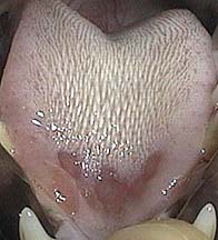
The tip of the tongue of this pet has an ulceration due to kidney disease
Blindness can occur due to the high blood pressure (hypertension) that develops as a consequence of CRF. We start therapy when the systolic blood pressure (BP) consistently exceeds 160 mm Hg.
Diagnosis
Since the symptoms of kidney disease mimic the symptoms of other diseases, a thorough approach is needed to differentiate them. In every disease we encounter we follow the tenet’s of the diagnostic approach to ensure that we make an accurate diagnosis, and also so that we do not overlook some of the other diseases that are also encountered in pets that have renal disease. Unfortunately, it is difficult to diagnose acute renal failure early in the course of disease.
Signalment
Kidney disease can occur at any age. If it occurs at a young age we tend to think more of toxins, cysts, and trauma. The most common form of kidney disease, CRF, occurs mostly in older pets.
Several feline breeds are prone to getting CRF as they age:
Persian
Abyssinian
Burmese
Maine Coon
Russian Blue
Certain canine breeds are also prone to CRF:
Lhasa apso
Basenji
Samoyed
History
Kidney disease is suspected in any pet that has some of the symptoms described above, especially PU/PD. The recent administration of medication, a recent bout of a disease, the changing of antifreeze, especially in the fall , and recent administration of anesthesia, are all helpful clues.
Pets that have other diseases that can affect the kidneys, notably heart disease, and hyperthyroidism, alert us to the potential for kidney disease.
Physical Exam
Symptoms noted during a physical exam depend on what caused the kidney’s to fail, how long the disease process has been present, and whether a pet has the acute form or chronic form of the disease.
Physical exam findings might include:
Pale gums due to anemia. You can check for pale games at home. Our Learning Center shows you how.
Dehydration
Small and irregular kidneys upon abdominal palpation if CRF is present
Large or nodular kidneys if a cyst or cancer is present
Underweight
Enlarged lymph nodes
Dilated or uneven pupils
Weakness
Ventro-flexion of the head and neck
Diagnostic Tests
Kidney disease can only be diagnosed with appropriate tests. As a general rule, we recommend screening for kidney disease by running a blood panel and a urinalysis on all pets greater than 8 years of age.
We also screen for other diseases, notably liver disease, sugar diabetes, and hyperthyroidism, on this blood panel due to their prevalence in older pets.
The most accurate test to assess kidney function is called the Glomerular Filtration Rate (GFR). You learned about the glomerulus in the anatomy section above. Unfortunately, due to the time and costs involved with running this tests, animal reference labs do not offer it. Instead we rely upon indirect assessment of renal function using the tests of Blood Urea Nitrogen (BUN), and Creatinine. Unfortunately, these tests are not elevated in the bloodstream until the majority of kidney function is lost.
To help, we look for biomarkers of kidney disease, which you will learn about in more detail. The primary one that is used is called SDMA. It is explained in more detail below.
New biomarkers are on the horizon, and as time goes on we will be able to assess their sensitivity (do they detect a kidney problem when it is present) and specificity (if they are elevated is it truly a kidney problem and not a problem with some other organ). These new biomarkers are (get ready for some tongue twisters when you pronounce them):
Urinary NGAL- Neutrophil gelatinase-associated lipocalin
Urinary GGT- gamma glutamyl transpeptidase
Immunoglobulins A, G, M
C-reactive protein
Thromboxane B2
Transferrin
RBP- retinol binding protein
Cystatins B and C
THP- Tamm-Horsfall protein
NAG- (N-acetyl beta-D-glycosaminidase)
KIM-1 (kidney injury molecule 1)
clusterin
F2- isoprostanes
Lymph node biopsy
Peripheral lymph nodes can be palpated in numerous locations. They can enlarge for several reasons, one of the more important ones is cancer. If they are enlarged and significant disease process is suspected, then one of them is biopsied (example to follow).
Blood Panel
An important tool in the diagnosis of kidney disease is a blood panel. This blood panel consists of a Complete Blood Count (CBC) and Biochemistry Panel (BCP). We look for abnormalities in several specific tests:
CBC- Complete Blood Count
This test checks the red and white blood cells. It is not uncommon for a pet with chronic kidney disease to have anemia. This anemia needs to be addressed.
The kidneys produce a hormone called erythropoietin that stimulates the bone marrow to produce red blood cells. Anemia occurs in kidney disease due to inadequate levels of erythropoietin, shortened survival time of red blood cells in general, bleeding in the stomach or intestines, and the effects of uremic toxins on parathyroid hormone. Pets that are dehydrated might not show anemia on a blood sample until they are rehydrated.
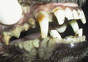
This dog has white gums in addition to the severe dental disease that is present. The white gums are due to anemia from CRF.
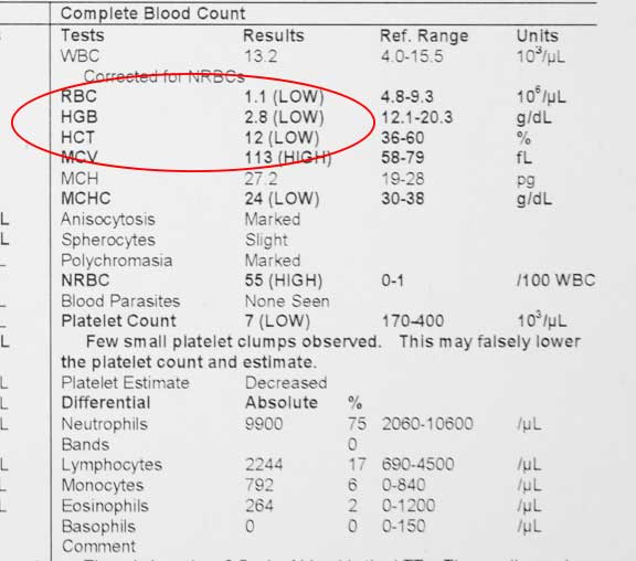
The next three blood panels show mild, then moderate, then severe anemia. Any one of these can occur in kidney disease.

Anemia is noted in this pet by the low RBC (Red Blood Cells), HGB (Hemoglobin) and low HCT (Hematocrit). This is mild, and might be correctable at this point.

This is moderate anemia
This pet has a severe anemia, and needs a blood transfusion if it is to survive
BCP (Biochemistry Panel)
There are many tests on a BCP, the following are the ones that relate to kidney disease.
BUN (Blood Urea Nitrogen)
The BUN is usually elevated in pets with kidney disease. This is called uremia. The nutrition of your pet can influence this.
BUN can also elevate in dehydrated pets and in pets with an obstructed urethra causing an inability to urinate. These are called pre-renal and post-renal uremia respectively.
If a urinary obstruction is the cause of an elevated BUN (post renal-uremia), the BUN levels tend to be extremely high. If dehydration is the cause of the elevated BUN (pre-renal uremia), then the values do not tend to be as high. The BUN must be interpreted in conjunction with a urine test called specific gravity to know if the BUN is elevated due to kidney disease or dehydration.
A low BUN is important in Liver disease.
Creatinine
It is the most widely used way to diagnose kidney disease, and is more reliable than BUN, since factors like dehydration are not as influential on creatinine as they are on BUN. This test is also a good early indicator of kidney disease even when normal, if the trend in values is increasing. This is not as accurate a market of kidney disease in pets that have lost weight and have decreased muscle mass (sarcopenia). Upon physical exam they will have a body condition score of less than 5/9.
This test does vary between individuals, so the normal reference ranges are a starting point. Trends should be monitored, using the same laboratory or the same machine. Since each pet might be different running this test early in your pet’s life when their is minimal chance of renal disease can help determine what is normal for your dog.
This again emphasizes the importance of yearly wellness exams as your pet ages. If the creatinine is going up, even if in the normal range, we might start treating for a kidney problem with Hill k/d Early Support. This increase in creatinine is part of the IRIS staging talked about at the beginning of this page.
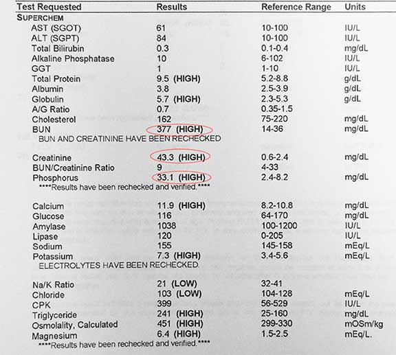
The BUN and creatinine are both elevated it is called azotemia. Unfortunately, by the time azotemia is present we have lost between 66% – 75% of the nephrons. So in reality, if they are elevated we are already in a hole. This is why the IRIS system was developed, and also why the SDMA test was developed, which you will learn about soon.
Phosphorous
In the more advanced stages of kidney disease the phosphorous levels elevate. When this happens the prognosis is not good. The Hill’s kidney foods have limited phosphorous for this reason.
Amylase
This is an enzyme produced by the pancreas to aid in the digestion of carbohydrates. It is excreted by the kidneys, so an excess in the bloodstream could indicate kidney disease.
The following four blood panels are from pets that have mild, moderate, severe, and terminal kidney disease. We want to catch this long before it even becomes mild.
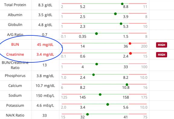
Even though we want to put a dog or cat on an appropriate K/D long before this, the azotemia on this patient is potentially treatable
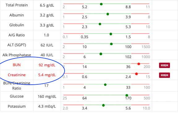
This moderate to severed kidney disease might not be treatable
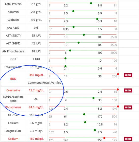
This severe azotemia, with a phosphorus that is 3X normal, is probably not treatable and euthanasia needs to be discussed
This pet has terminal kidney disease and is in the act of dying. It is surprising it is still alive with a creatinine that is 18X normal
SDMA
Serum symmetric dimethlyarginine is a new kidney marker that might aid in early diagnosis of this malady. It is an amino acid (arginine) that is excreted by the kidneys. Less than 14 is considered normal. An advantage over creatinine is the fact that it is not influenced by muscle mass, and a pet that is underweight does not have an effect on this test like it does with creatinine.
Increases in this test occur prior to increases in serum creatinine if a normal creatinine is considered 2.4 or less. SDMA might not be accurate in pets with Diabetes Mellitus, neoplasia (cancer like lymphoma), and kidney stones (nephrolithiasis). The most important here, like other tests, is to follow the trends, and even if normal, note if they are increasing.
FGF-23
This test monitors a compound called fibroblast growth factor 23. It is a hormone that increases when the BUN and creatinine are elevated. It helps detect excess phosphorous earlier than a blood panel monitoring phosphorous can.
Urinalysis
This is also an important tool in the diagnosis of kidney disease and another early indicator of kidney disease along with creatine. Changes in several parameters could indicate kidney disease:
Specific Gravity (S.G.)
The ability of the kidneys to dilute and concentrate the urine is an important parameter to monitor. Water has a specific gravity of 1.000. A pet with kidney failure has a specific gravity of between 1.008-1.012. A specific gravity in this abnormal range is called isosthenuria. In dogs with normal kidney function the S.G. should be greater than 1.025.
This number is interpreted in conjunction with the BUN to help determine if the elevation in BUN is due to dehydration or kidney disease. To complicate things further, dehydration and kidney disease can occur simultaneously. Also, as mentioned above, liver disease, a common problem in older pets, can also be an influence. To be accurate the specific gravity should be checked immediately after obtaining a urine sample.
Protein
Excess protein in the urine, called proteinuria, is a common finding in CRF. It can also occur in glomerulonephritis, pyelonephritis and amyloidosis.
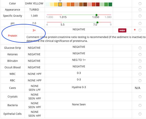
A urine protein of 3+, with a Specific Gravity of 1.049, and no sediment, is significant, and warrants further testing.
There is evidence to suggest that urine protein:creatinine ratio can be a predictor of survival time. Pets with a ratio < 0.5 tend to live significantly longer than pets with a ratio > 0.5.
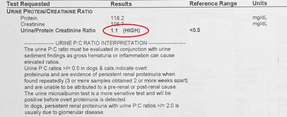
This pet as a high ratio, indicative of kidney disease
Cells
Specific types of cells, called casts, can also be an indication of kidney disease. The urine sample in the pet above with the 3+ protein has none of these cells.
Urine Culture and Sensitivity
If pyelonephritis is suspected, or bacteria are noted in the urinalysis, the urine should be cultured to determine which bacteria if any is present. If a bacteria is grown out then the appropriate antibiotic should be used for 4-6 weeks. A urine culture with an E. coli bacteria that was resistant to several routine antibiotics was shown earlier in this page.
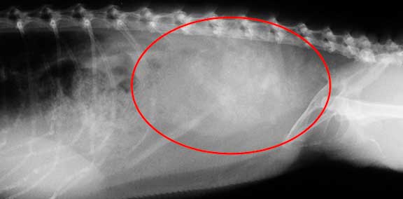
Radiography
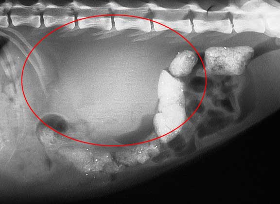
Radiography can be very helpful in the diagnosis of kidney disease. It allows us to visualize the kidneys, check for stones in the urinary system, look for calcification that might go along with kidney disease, and also look at other organs that commonly have a problem as pets age.
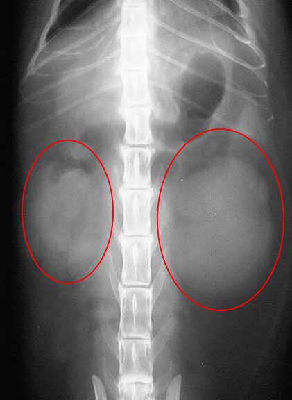
These kidneys have a normal size and shape. Use this for comparison purposes as you look at the other radiographs.
The two overlapping right and left kidneys are circled, although they appear only as one. Can you see both of them? The right kidney in mammals is more forward than the left.
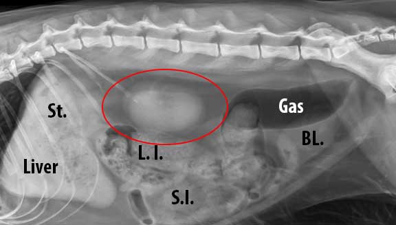
St.- Stomach
L. I.- Large intestine
S. I.- Small Intestine
Bl. – Urinary bladder

This is the radiograph of a pet with normal kidney’s that is lying on its right side. You can see the right kidney (RK) more forward in the abdomen compared to the left kidney (LK). The area of the 2 kidney’s that overlaps is more whitish in nature. This is called summation, and there is nothing wrong with these kidneys.
This is the radiographic of a pet with renal lymphosarcoma (malignant cancer). The diseased kidney is the large white circular area in the center of this view. It is pushing the large intestine down. We have a Kidney Cancer page showing how this is diagnosed and treated in a young dog.
The left kidney is larger than the right in this view, called a V-D (ventrodorsal) view of the abdomen, a potential sign of cancer
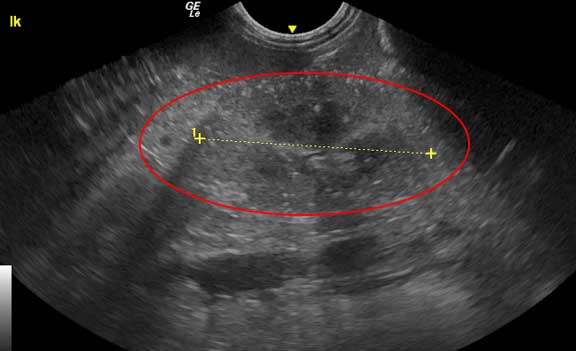
Ultrasound
A very valuable tool in the diagnosis of kidney disease is ultrasound. It allows us to look at the ureters and bladder, internal anatomy of the kidney, measure kidney size, and take a biopsy for an accurate diagnosis. In many cases the use of ultrasound precludes us from having to perform an exploratory surgery.

This is an ultrasound of the right kidney, which is being measured to determine its size
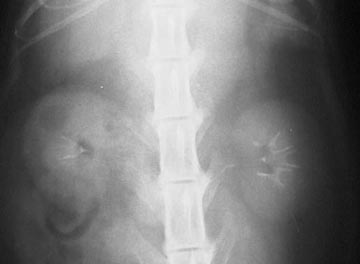
Excretory Urogram
This special test, also know as an IVP (intravenous pyelogram) gives us significant information about the renal system. It has to be used carefully if ARF or CRF is suspected because it can exacerbate the problem. A radiopaque dye is injected into the bloodstream and radiographs are taken of the dye as it passes through the kidneys, ureters, and bladder.
Laparotomy
Exploratory surgery (laparotomy or celiotomy) is frequently used as an aid in the diagnosis and treatment of renal disease, especially cancer. We use this option when we feel that ultrasound will not be advantageous.
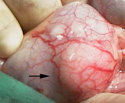
The arrow points to a lump on the surface of a kidney. It was caused by cancer that spread from the stomach.
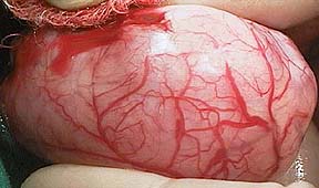
A section of the lump was biopsied during surgery to determine the cause. The tremendous blood supply to the capsule that surrounds kidneys can easily be visualized. Even though this capsular blood supply is extensive, it pales in comparison to the amount of blood that flows into and out of the kidneys through the renal artery and veins.
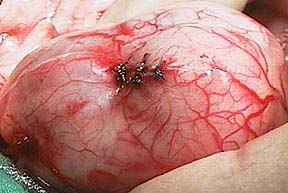
Three sutures were placed in the kidney capsule to control the bleeding that occurred at the biopsy site
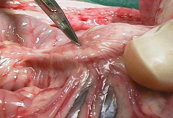
A biopsy of a lymph node (called cranial mesenteric) located in the center of the abdomen was also obtained. This helps us determine if the cancer has spread.

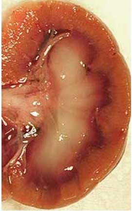
This is what cancer looks like inside a kidney that has been cut open. The arrow points to the problem on the left side. Compare this to the normal kidney architecture that goes from around 2 PM to 8 PM.
Treatment
Acute Renal Failure (ARF)
This form of renal disease needs immediate and aggressive treatment to prevent death. Any drug suspected of causing the problem is stopped immediately, and underlying problems are addressed,
Ethylene glycol (antifreeze) poisoning is an example of ARF. Cats that have a urinary obstruction need to be unblocked immediately. If not, excess potassium in the bloodstream (hyperkalemia) can cause death to to its affects on the heart.
Fluid therapy is critical, and consists of saline and dextrose solutions initially. Fluid therapy corrects fluid and electrolyte imbalances, increases the blood flow to the kidneys, and starts the process of diuresis.
Pets need to maintain their caloric input in order to minimize the metabolism of protein for their caloric needs. Metabolizing excess of amounts of protein will increase uremia, causing a further deterioration in condition.
Pets that are still not urinating after this initial fluid therapy are given Lasix or mannitol. Excess potassium (hyperkalemia) is a common finding in ARF. If mild, fluid therapy alone should correct the problem. If severe, regular insulin and sodium bicarbonate are used.
Pets with ARF are sensitive to ulcers and infections, so treatment for these problems is sometimes initiated.
Pets that have heart disease are sensitive to IV fluids because excess amounts can cause an overload to the lungs called pulmonary edema. These pets pose a dilemma. If we do not give them enough fluids the kidney problem will worsen. If we give enough fluids to help flush the waste products out of the bloodstream, these same fluids might cause pulmonary edema.
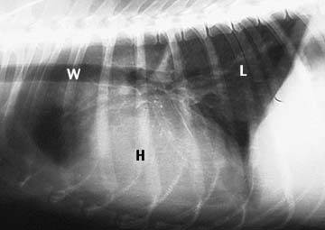
This radiograph is of the chest of a normal dog. The heart (H), windpipe (W), and lungs (L) are labeled. The lungs are black because they are filled with air. This is how normal lungs look on a radiograph.
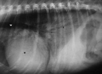
This dog has pulmonary edema. The air filled lungs are no longer black, they are white from the fluid that has built up. This is a very serious condition.
Chronic Renal Failure (CRF)
This is the version of kidney disease we encounter most often. The prognosis is guarded, and depends significantly on how long the disease process has been present along with your pet’s age. Pets (usually geriatric) that have other diseases that are common at this age can make this difficult to treat if not caught early enough.
Many pets (especially cats) that are brought to our hospital have CRF that has progressed to the point where the problem has become similar to ARF. These pets need to be hospitalized and put on intravenous fluids almost continuously to get them over this acute phase.
We will closely monitor their BUN and creatinine before therapy is instituted and during hospitalization, to ascertain if their kidneys are responding to fluid therapy. If the BUN and creatinine do not drop significantly after 24-48 hours of intravenous fluids then the prognosis for recovery is poor.
Many treatments have been advocated to help minimize the symptoms of CRF (also called the uremic syndrome). None of them can cure the problem, and not all of them have proven to work, so it is important that we tailor make each pet’s therapy to its individual needs.
In addition, indiscriminate use of medication to treat a perceived problem can make the kidney disease worse. This applies to almost every drug, since the kidneys are so intimately involved in the metabolism of drugs. The medical axiom of “first do no harm” applies directly to kidney disease.
Medical management of CRF needs to address the following:
- Protein and phosphorous regulation
- XS protein in the urine
- High blood pressure
- Anemia
- Dehydration and electrolyte imbalance
- Low pH in the bloodstream
- Stomach and intestinal ulcers
Fresh water should be available at all times for your dog. You should empty and fill the water bowl at least 3 times per day to help stimulate drinking. Place water bowls in several locations around the house for your older pet, especially in multi pet households. Undue stress should also be minimized at all times also.
If you are a cat owner please read our page on Stress In Cats to help understand this important aspect of feline health.
Diet
Early intervention with a kidney diet is essential to prevent this chronic problem from progressing faster than it need be. Pets with CRF need to be fed a diet that has limited amount of high quality protein. Less protein in the diet leads to less work the kidneys have to perform by removing the nitrogenous waste products that are the end result of protein metabolism. Protein is vital to all bodily functions and can not be indiscriminately limited. As a matter of fact, if protein restriction is not implemented carefully it can make the uremic syndrome worse.
High quality protein means that it contains more essential amino acids, which are those the body cannot produce and must be obtained in the diet. This was talked about at the beginning of this page, and is of crucial importance in treatment.
These foods also need to be limited in phosphorus. This is not the case with many pet foods since taking phosphorus out is a more costly endeavor for a manufacturer. The food also needs to have an adequate amount of potassium, sodium, and omega- 3 fatty acids, water soluble vitamins, and antioxidants, and they need to be in proper proportion to each other.
Mixing this new food in partially with the regular diet and heating it up a little (for the canned food) in the microwave helps. Adding a small amount of a tasty fluid like clam juice can make it taste better.
The use of appetite stimulants, which you will learn about below, are beneficial here. It is important to keep your pet eating so that it does not lose muscle mass, another one of our IRIS staging parameters.
Water soluble vitamins (ex.- B-complex vitamins) are easily depleted in a pet that has PU/PD. Supplementation can be helpful.
Phosphorous lowering medication
Pets with CRF might have an increase in their phosphorous levels as the disease progresses. Performing the FGF-23 can be helpful in addressing this phosphorous problem before it progresses. This excess phosphorous can add to the anemia that is common with CRF. It will also dramatically influence calcium metabolism in the body through a hormone called parathyroid hormone. The end result will be painful calcium deposits in the bones and internal organs, including the kidneys. This will also add to the scarring and add to the progression of CKD.
As already mentioned, K/D©is restricted in phosphorous, and should be used in combination with phosphorous binding medication. The phosphorus binding medication we use, which always needs to be given with food, is called Epakatin® by Vetoquinol©.
Potassium increasing medication
Pets with CRF will have a decrease in their potassium levels as the disease progresses. This does not always show up on a blood panel. Using oral supplements and adding additional potassium to fluids helps counteract this problem. Oral potassium is called Renal K+®, and it comes in a paste for easier administration.
Urine protein reducing medication
Reducing protein in the urine is believed to help slow down the progression of the disease. ACE (Angiotensin Converting Enzyme) inhibitors (ex- Enalapril) or ARB’s (Angiotensin Receptor Blockers) are used when the urine protein:creatine ratio is greater than 0.5 in the dog and greater than 0.4 in the cat. These drugs are given for 30 days and then the urine is checked to see if there is either a 50% reduction from the original test, or if the ratio is below 0.5 in the dog and 0.4 in the cat.
Anemia fighting medication- Darbopoentin (Aranesp)
One of the long term affects of CRF is anemia due to a lack of erythropoietin secretion from the kidneys. This anemia can occur in up to 2/3rds of the pets with kidney disease. This hormone can be supplemented to help minimize anemia. In many pets that are treated with K/D food and fluids, all that is needed to counteract this anemia is a little time for the uremic waste products to diminish in the bloodstream. Giving these pets extra iron in the form of Iron Dextran injections every 7-10 days initially is beneficial.
A drug with conditional FDA approval to treat anemia in cats is called Varenzin-CA1 (molidustat oral suspension) from Elanco. This drugs stimulates the kidneys to produce erythropoietin. One of our doctors might recommend it when it has proven to be both effective and safe in a clinical setting when used on many cats.
If a dog has anemia from CRF it needs to be monitored. We have an easy to perform and inexpensive in-house blood sample that only requires a few drops of blood from your pet. It is called HCT/T.P. This stands for Hematocrit and Total Protein. The HCT gives us a percentage or red blood cells. It is a part of all of the CBC’s presented earlier in this page.
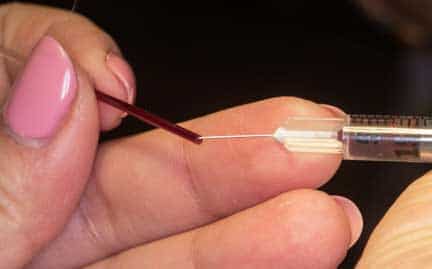
So little blood is needed that we can take blood from tiny birds and reptiles this way
Fluids
One of the most important treatments for CRF is the administration of supplemental fluids. Whenever we tell people their pet with kidney disease needs fluids they commonly respond “its OK, he/she already drinks a lot of water”. Unfortunately, this excess drinking of water is a result of kidney disease, and not a sign that the pet is drinking adequate amounts of water.
Some pets are not good drinkers, and need additional water to what they are already drinking. Feeding canned food compared to dry food is a big help here, so try that if you are not already doing this. Adding nutrient enriched supplements to your cat’s drinking water can help, along with keep the water fresh and even have a system of constantly running water. Whatever it takes, get you cat to take in more water.
If your pet is hospitalized we will give them intravenously (IV) because of greater effectiveness and accuracy. If your pet responds to IV fluids during its hospitalization we will initiate the use of subcutaneous (SQ.) fluids at home on a daily basis.
This area of home treatment is so important that we have devoted a complete page to its use. Please click here to learn about the proper technique, then return to this section for more treatment options.
We commonly add B-complex vitamins to the fluid bag since these water soluble vitamins are excreted the more we give supplemental fluids. Feeding a food designed for kidney disease like K/D will also help minimize the depletion of water soluble vitamins.
Blood pressure medications
Earlier in this page you saw how we take a blood pressure reading.
Hypertension is a common occurrence as the disease progresses. Any systemic blood pressure of 160 mm or more should be treated. Blood pressure lowering medications like Norvasc (calcium channel blocker) and Enalapril (ACE inhibitor) will help counteract this problem. All pets initially diagnosed with CRF should have a blood pressure taken. It should be rechecked at least every 6 months.
Some pets might respond to ACE inhibitors to decrease the protein in their urine. Further studies are needed in this area to determine efficacy.
Anti-ulcer medication
Some pets with CRF don’t eat well because of nausea due to excess hormone secretion in the stomach. Tagamet (famotidine) and Prilosec (omeprazole) are used to counteract this problem. If we suspect an ulcer in the stomach due to the toxins that have built up we mights use these medications also.
Anti-vomiting medication
Vomiting is a common problem in pets with CRF. It occurs as a result of uremic toxin buildup in the bloodstream and alterations in hormones that regulate gastric secretions. This is a sign of how sick they are, and it also precludes them from getting proper nutrition, which is crucial in the treatment.
Vomiting will cause dehydration, leading to a decreased flow of blood to the kidneys (decreased GFR) and an increase in azotemia.
We use a drug called Cerenia (maropitant) to alleviate vomiting symptoms. There is an injectable form of this drug used in an acute case, and an oral version for long term use.
Antibiotics
Animals weakened by kidney disease are more susceptible to infection. These pets are commonly older and have significant dental disease. Antibiotics help them fight off infections. The antibiotic dose might have to be adjusted since many of them are removed from the body by the kidneys.
Pets with CKD commonly have urinary tract infections due to bacteria, so a urine culture and sensitivity is needed in these pets to see if this problem is present.
Appetite stimulant medication
Pets with CKD do not eat well for many reasons. Getting them to eat is crucial, and some of them need appetite stimulants. The most effect one we use is called Mirtazapine. Mirtazapine comes in a topical version if you cat is not eating well.
Elura from Eland should be fed to your cat if it is not eating well. Elura mimics the action of a hormone called gherlin that stimulates the appetite.
Probiotics
This supplement digests non-protein nitrogen in the intestines, minimizes BUN and creatinine levels, so there is less work for a diseased kidney.
Feeding Tubes
This overlooked and effective treatment helps dramatically for pets with CKD that are not eating well, are vomiting, and are difficult to medicate orally and with SQ fluids. We have a detailed page on feeding tubes.
Miscellaneous treatments
There are other supplements and medications used in CRF that might be of some benefit, although this is unproven. As long as they do not cause the problem to worsen they might be worth trying.
Anabolic steroids– They are also used in older pets for arthritis and appetite stimulation with good success. They might help counteract the affects of anemia, although this can not be relied upon. DecaDurabolin® is the one we used most frequently. Drugs like this are no longer readily available.
Calcitriol– It is postulated that some of the symptoms of CRF are the result of elevated levels of parathyroid hormone. The IRIS does not recommend the use of this due to a lack of evidence that it helps.
Kidney Transplantation
At some select veterinary universities (University of Georgia and University of Pennsylvania) a new kidney can be transplanted into a cat. Success rates is over 75%, and almost half of the patients that receive a kidney transplant survive at least 3 years after the surgery. One requirement of the program is that the donor cat needs to be adopted.
In addition to chronic kidney disease, other common causes for a cat to need a kidney transplant is eating lilies, antifreeze (ethylene glycol) toxicity, polycystic kidney disease (PKD), pyelonephritis, and renal amyloidosis.
Pre-operative screening is important to make sure the donor and recipient cat are physiologically capable of this, and the owner understands the emotional and financial commitment needed. This includes the need for lifetime immunosuppressive drugs like cyclosporine, the need for diagnostic follow up tests, and the potential for rejection of the kidney. This rejection can occur in up to 25% of the patients. Diseases like diabetes mellitus and cancer can also be associated with kidney transplants in cats.
This specialized surgery can cost upwards of $10,000.

This is Skipper with his wonderful mom in for special testing before he goes to Georgia for his kidney transplant

This is a picture of him during the surgery. The organs are labeled below for your understanding.
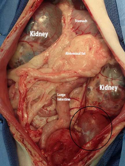
The old, swollen, and diseased kidneys are labeled. The new kidney is circled

Skipper returned several months later feeling much better, and with his new brother!
Prognosis
Pets presented with renal disease, whether ARF or CRF, carry a guarded prognosis. This emphasizes the need to put your 8 year old cat on Hill’s c/d before this common problem progresses.
If your pet is hospitalized with CRF we will closely monitor its blood panel, paying special attention to BUN, creatinine, and phosphorous. If the excess levels of these tests decrease dramatically during hospitalization, and your pet improves clinically, then the use of k/d food, medications and SQ fluids at home are usually advantageous.
All pets that have been diagnosed with CRF should have a blood panel, a urinalysis, blood pressure check, and physical exam performed every 3-6 months. This disease will progress, and other diseases might present themselves, so this type of monitoring is crucial for a good quality of life.
Return to the Diseases page.