
Introduction
Dental disease is prevalent in almost every one of the dogs and cats we examine. This page has a large amount of information that will inform you of this serious and overlooked problem. Please set aside the time to fully understand it due to its importance regarding your pet’s quality of life.
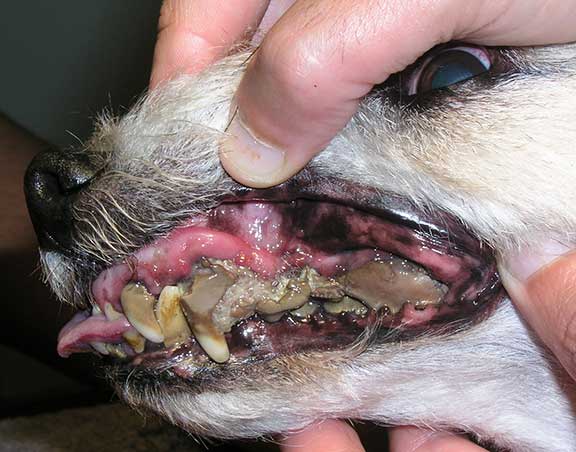
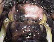
We see too many pets presented in a state similar to this. The periodontal disease in this dog has progressed so far that it is systemically ill, and in danger of internal organ failure and a spontaneous jaw fracture. Imagine how this dog feels. Not only that, for a species with such an acute sense of smell the odor must be nauseating.
Why is it overlooked? Most people do not readily look in their pets’ mouths. They also don’t understand the importance of dental care in regards to bacteria, pain, and quality of life. That is what this page is about.
Also, there is this irrational fear of anesthesia based on old fashioned notions about the risk. This is addressed on this page, starting with non-anesthetic dentistry.
People sometimes wonder why pet’s need their teeth professionally cleaned by us, and then brushed by you, when they have memories of growing up with dogs and cats and never doing this. It doesn’t take much to answer this question. Pets nowadays eat diets that makes them prone to plaque. They also live longer, and just like in people, are more prone to disease as time goes on.
More importantly, we did not have the knowledge decades ago to understand how dogs and cats lived lives of chronic pain because we did not know, or could not diagnose, the periodontal disease that is occurring below the gum line.
With the advent of digital radiography, and our current body of knowledge, we realize that we did not treat dental disease anywhere near as thoroughly as needed. This ignorance lead to poor quality (painful) and shortened lives for our pets.
Dental disease is a treatable and preventable problem, and since your pet cannot tell you how it feels, it is up to all of us, as members of your pet’s health care team, to address this problem. Most people wait too long to get their pets teeth cleaned professionally.
As we increase our knowledge of animal health we realize that proper dental care does not just make your pet’s breath smell better; it is mandatory for your pet’s long term quality of life.
This problem is prevalent in many species, even animals like ferrets.

The three areas of gingivitis in this ferret are circled in blue
Comprehensive Information on Dental Disease
Our dental disease in animals page is comprehensive, and is broken down into 13 major sections:
- Examples of dental disease in dogs and cats
- Prevention
- Treatment with non-anesthetic dentistry
- Tooth anatomy
- Biofilms (bacteria) that are a major part of dental disease
- Symptoms of dental disease
- Stages of dental disease
- Internal organ problems associated with dental disease
- Diagnosis of dental disease
- Anesthesia used for dentistry
- Professional cleaning
- Congenital abnormalities
- Miscellaneous dental problems
We follow the principles of the American Veterinary Dental College in treating pets with this serious problem.
Oral hygiene is one of the most overlooked areas of medical care for animals. Far too many pets come to us with advanced dental disease, requiring anesthesia, x-rays, and the removal of rotten and painful teeth. Some of these pets are systemically ill as the billions (yes billions) of bacteria in their mouths enter the bloodstream through diseased gums and seriously infect important organs like the liver, kidney, and heart
The following pictures illustrate what happens when dental disease is not properly addressed. The pictures are from a dog under anesthesia to have its teeth cleaned.

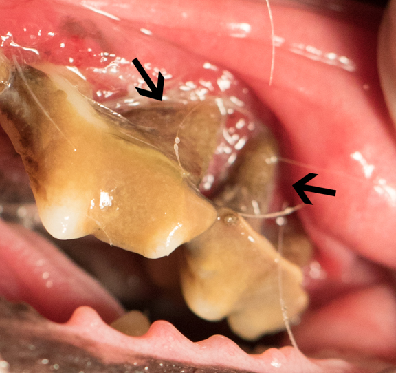
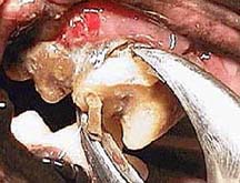
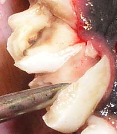
This is the crux of the problem. When you look at the teeth, and see some tartar, it doesn’t seem like too much of a problem. Just scrape the tartar off and the teeth will look and feel better, and your pet’s breath will smell better. Job done right?

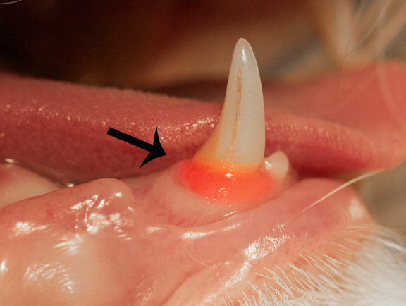
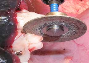
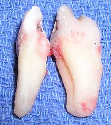
Here is the same tooth as it is being removed while this pet is under anesthesia. The root on the right is rotten, compare it to the normal root on the left. We did not know this pet had a rotten and painful root until we probed it and took radiographs. If this tooth had only been cleaned of tartar and not removed, this pet would have had a painful tooth indefinitely.

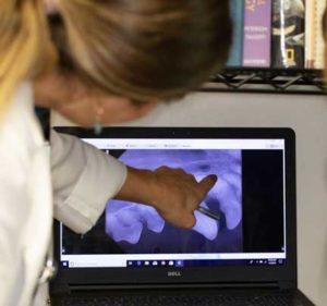
We were able to confirm this rotten root by taking digital dental X-rays
This is a video of a different dog having its teeth cleaned under anesthesia. When our doctor examined the canine tooth it was found to be infected, with pus coming out of the gums. This would never had been found if we were not thorough and checking for this. This dog would have had a painful mouth indefinitely if we had not treated it. Just as importantly, the large amount of bacteria that is entering the bloodstream on a chronic basis can cause damage to the internal organs.
You can see the pus on the probe as it is moved around the base of the canine tooth
Cats get a unique dental problem, called neck lesions (also called FORL- Feline Odontoclastic Resportive Lesions), that are painful. When we encounter these problems we need to remove the bad tooth.
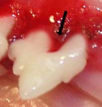
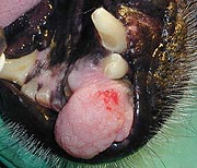
What seems like just a red gum is actually the painful condition of the root called FORL
Here is another example of a potential FORL lesion
The short video below is an example of how painful this is. This cat is completely anesthetized, yet when we gently touch its premolar teeth with a probe, it moves its jaw in obvious pain. This is the pain it perceives while under general anesthesia, think of how it felt when it was awake prior to treating it.
It is the periodontal disease that is occurring out of sight and below the gum line that causes the most problem. This is the area we thoroughly need to address when we clean a pets teeth under anesthesia.
We take this problem seriously, and spend considerable time caring for pets with dental disease. Many dogs and cats are brought to us in advanced states of dental disease. Their teeth are infected and rotting out, and they have tremendous odor (severe halitosis) from the infection.
For animals that have such keen senses of smell, this chronic odor is very irritating. The stress on their internal organs due to the tremendous infections in their mouths can cause problems with the liver, kidneys, and heart valves.
Dogs and cats with advanced dental disease need to be anesthetized, given a thorough oral exam, and have dental radiographs.

They are closely monitored during the procedure, and all their dental problems are taken care of. This takes considerable time and effort on our staff as we carefully assess every tooth.
The following information on dental disease is very thorough. We have a summary page on dental disease if that suits your needs better.
Prevention
Prevention is the key. We will talk about this prevention when you bring your pet to us at a young age. This way its never suffers painful and infected teeth, and will not need to go under anesthesia for tooth removal.
Teeth cleaning should be considered a preventive measure, not a way to treat an infected tooth or gingivitis that is already present. Good dental care revolves around the control of bacteria under the gum line where it is not visible. We will teach you how to prevent it and how to treat it.
Start with learning how to brush your pet’s teeth. If started at an early age this “bonding time” is an enjoyable time for all. When your dog or cat is young, get it used to your hands around its mouth by petting it on the head and face gently, telling your pet how good he is in a soothing tone.
Eventually get him used to your fingers being gently placed in his mouth and rubbing his gums. If you do it slowly and follow your pet’s reaction, you can make this a fun game for all. At the end of this page on dental disease there is more information on prevention.
When your puppy or kitten still has its baby teeth use our dental wipes to get your pet used to the procedure. By going slowly most puppies and kittens respond positively to the attention. You can dip a cotton-tipped applicator in tuna juice and use this to rub kitten gums.
Start brushing the teeth when the adult teeth are in, which is around 5 months of age. We can tell you when to start if you are not sure, and show you how to do it.
Yes, you can brush your pet’s teeth, and yes, you can make it an enjoyable experience for both of you! Bring your pet in and we will show you how. If you start when your pet is young it will be much easier. This can be started when you do your puppy behavior training.

We have special dental kits for dogs and cats that make the process easier and more effective. They do not like the feel and taste of the toothpaste we use in our mouths, so do not use that.
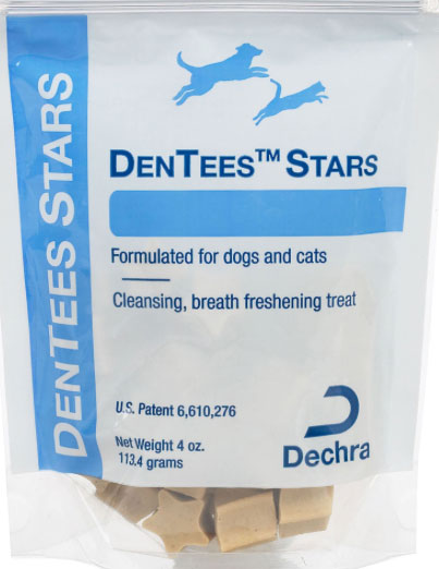
If brushing your pet’s teeth is not an option, we have a multitude of products to help replace brushing. They are designed to aid in slowing down plaque buildup, which is the start of dental disease. They are not as effective as brushing, but are better than doing nothing.
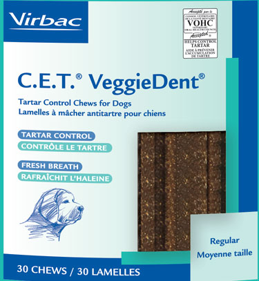

There is also a food called T/D (tartar diet) made by Hill’s which is a significant help in preventing dental disease. It comes in many different kibble sizes for different sized pets.We highly recommend it, especially for the small breed dogs that might be fussy about getting their teeth brushed.
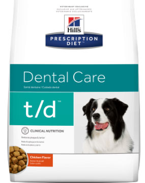
This food is unconditionally guaranteed, and can be returned for any reason
Non-anesthetic Dentistry
If you use some of these products, and have your pet’s teeth cleaned by us without anesthesia (it’s called non-anesthetic dental (NAD), there is much less chance your pet will need to be anesthetized to take care of painful teeth. All of this needs to be started early in your pet’s life, and long before dental disease sets in. When your dog completes its vaccine series and gets spayed or neutered is when we will start talking about implementing this.
Having your pet’s teeth cleaned professionally without anesthesia, when it is young and dental disease has not set in, is the best way to prevent dental disease. After they are cleaned this way you need to brush their teeth, until their next cleaning in 6 months.

We utilize the services of the most experienced and professional company in the business named Pet Dental Services
They are very skilled at what they do, and almost every pet does fine without any sedation
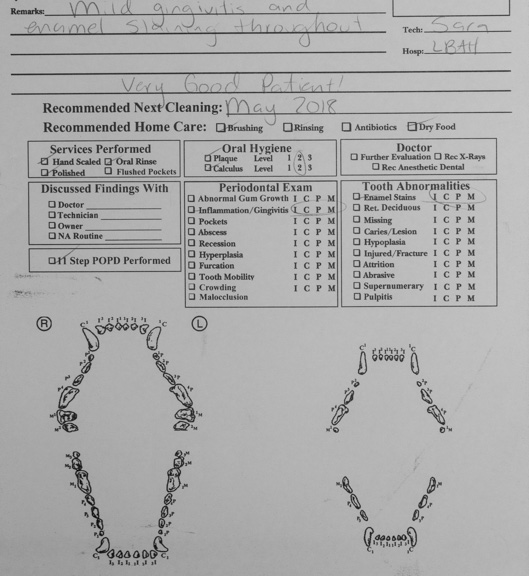
The non-anesthetic professional teeth cleaning by Pet Dental Services is thorough, and the person that cleans your pet’s teeth will go over a report like this for your pet.
The licensed technicians from Pet Dental Services perform non anesthetic teeth cleaning at our hospital on the 4th Monday and Wednesday of every month. You can make an appointment and wait while it is being performed, or you can drop your pet off and pick it up later. If you wait it takes about 30 minutes.
All legitimate non anesthetic dental people are licensed by the state of California to perform this procedure. This can only be performed legally in the state of California under the direct supervision of a licensed veterinarian, which is why our doctors are present, and review any important dental findings with you after they professionally clean your pet’s teeth. This law is obvious for your pet’s protection.
Many unscrupulous people perform this procedure because they tell you they are saving you money and fooling you into thinking they are actuallly doing something medical for your pet when they clean your pet’s teeth without the supervision of a veterinarian. This commonly happens at grooming shops and pet stores. They prey upon the irrational fear people have of anesthesia.
In reality, since they are not making a correct diagnosis, or doing a thorough job, all they are doing is setting up the stage for the bacteria that is under your pets gum line to to wreak havoc later on, necessitating an anesthetic dental. The teeth look nice, which means you have done something cosmetically nice for your pet, but you haven’t touched the medical problem.
Normal Tooth Anatomy & Development
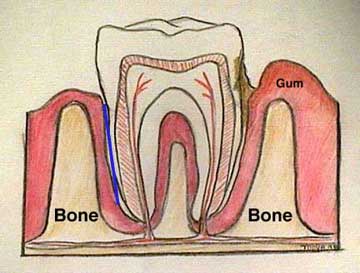
The diagram above illustrates some of the structures of the normal tooth. It also shows Stage III periodontal disease, which you will learn more about later. On the left side you can see the bone of the jaw and the blue periodontal ligament. It is this ligament that keeps the tooth attached to the bone in the socket. You can also see the blood supply and nerves to the tooth. They are the vertical finger-like projections in the center of the tooth.
On the right side we have illustrated what happens in gum disease. The brown area between the tooth and gum is tartar and its associated bacteria. Tartar by itself is inert, and does not cause dental disease. Removing it makes the teeth look better, but it does not address the primary problem. It is the bacteria surrounding and within this tartar that we are after. Notice how a significant amount of tartar is below the gumline, and thus cannot be seen. Also, notice how the gum is pulled away from the tooth leaving a pocket.
As the bacteria progresses further down the tooth, the gum is pulled further and further away, the jawbone literally erodes away, and the periodontal ligament can no longer hold the tooth in the socket. The tooth is painful, rots out, or is removed when we professionally clean the teeth. The bacteria that eventually causes this erosion enters the bloodstream and can cause disease in other organs. It is this bacteria below the gum line that is causing all the trouble, and is the bacteria we remove when we professionally clean the teeth.
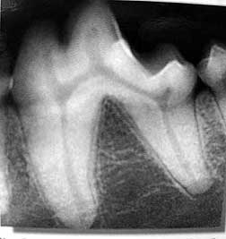
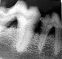
This radiograph of a tooth shows the same anatomy as above. We will show it again later when we show radiographs of diseased teeth. Notice how tightly the roots of the tooth fit into the healthy jaw bone. When we show you radiographs of diseased teeth later this jaw bone will be partially gone.
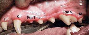
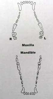
Dogs have 28 deciduous (temporary or baby) teeth and 42 permanent teeth. Anatomically they have 4 different types of teeth: Incisors (I), canines (C), premolars (Pm), and molars (M)
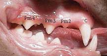
In comparison to dogs, cats have 26 deciduous teeth and 30 permanent teeth. They have the same types of teeth that dogs do, but in different proportions. They lack premolar #1 found in dogs due to a different evolutionary path.
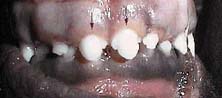
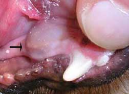
The deciduous teeth start being replaced by the permanent teeth (in this picture they are the 2 large central incisors marked by the arrows) at 4 months of age. The puppy teeth that were there were probably swallowed.
Dogs seldom have problems with teething, although they do tend to chew things during this period. It is advisable to supply them with synthetic bones for this purpose, or else some of your personal items might get recycled! By 8 months of age all the permanent teeth have appeared. Our Puppy Behavior page has more info on training with these bones.
Biofilms
Dental disease is all about bacteria. Due to the unique environment of the mouth, we measure mouth bacteria counts in the billions! Bacteria that adhere to the enamel of teeth colonize and begin synthesizing molecules, the most important of which are carbohydrates. These carbohydrates are sticky and act as a glue to attract more molecules on the teeth, eventually forming plaque.
As time goes on calcium carbonate deposits on the plaque, hardens, and then becomes calculus. This is the hard material deposited on teeth people sometimes call tartar.
Tartar is made up of calcium salts, food debris, bacteria and other organic matter. It is orange to brownish in color and although soft when deposited, it quickly hardens. It collects primarily on the cheek (buccal) side of the premolars and molars, although it can occur in any tooth.

This is tartar (plaque) on the teeth of a cat. If we get on top of this plaque now, by cleaning this tooth professionally with non-anesthetic dentistry, and use the prevention measures discussed earlier, we can prevent it from getting gingivitis, and all its associated problems.
Periodontal disease results when the bacteria at the center of this plaque move under the gumline. There are many different bacteria in the mouth that start the process of plaque development. Some are aerobic, and live off the rich oxygen supply in the mouth (can you guess why the mouth has a rich oxygen supply?).
As mentioned above, some of the bacteria in the plaque that migrate under the gumline go to an area of no oxygen, and are called anaerobic. The anaerobic ones tend to cause the most problem. Here is a list of some of their scientific names (warning-they are tongue twisters, so you better brush up on your Latin):
- Actinobacillus actinomycetemcomitans
- Bacteroides asaccharolyticus
- Fusobacterium nucleatum
- Eikenella corrodans
- Porphyromonas gingivalis
- Actinomyces viscous
These anaerobic bacteria cause an inflammatory reaction, and break down the periodontal ligament. The end result; the tooth rots out and the nerve is inflamed and painful. Also, as these bacteria invade deeper into the tooth cavity they reach the blood supply to the tooth and enter the bloodstream where they cause significant damage to the liver, kidney, and heart. It can even predispose pets to diabetes mellitus (sugar diabetes).
The problem does not end there when it comes to periodontal disease. It can also lead to spontaneous jaw fractures, deep seated bone infection, and cancer (neoplasia) at the affected tooth.
Since bacteria are the main culprit in periodontal disease, it makes sense that antibiotics will be used to treat the problem. Antibiotics are certainly no replacement for professional cleaning, but have a place in some select cases to help minimize the bacterial load and the halitosis.
How do you prevent these bacteria from starting the problem all over again after the teeth are professionally cleaned? Use the preventive care products mentioned at the beginning of this page, and get your pet’s cleaned professionally without anesthesia (non-anesthetic dental) every 6 months.
Symptoms of Dental Disease
Symptoms of dental disease can range from subtle to extreme. One of the most common symptoms is bad breath (halitosis). Sometimes a pet with dental disease will shy away or cry in pain when you touch it anywhere near its muzzle. Another symptom is a partial or complete inability to eat (anorexia).
A pet that has this problem may eagerly go to the food bowl, and either just look at the food or drop the food out of its mouth after only a few bites. Other pets might drool from one or both sides of the mouth, or paw at the mouth. Unfortunately, many pets are stoic (do not show outwards signs of pain when it exists on the inside), and do not show any symptoms until the problem is well entrenched, and the roots are rotting and painful.
Pawing at the mouth from dental disease
The important point to remember is the fact that once you notice any of these symptoms, your pet’s dental disease is already causing discomfort or pain, and even affecting other body organs. Therefore, it is important for you to be aware of the existence of this problem, to learn how to perform a basic oral exam at home, learn how to brush its teeth, and to bring your pet in for regular (every 6 months) dental exams by one of our veterinarians.
An exam every 6 months might seem like a lot to some people. Compared to the typical lifespan of a dog or cat, it is not very frequent. Your pet cannot tell you its mouth hurts, it is up to us, as a team, to ensure that this inevitable problem is properly monitored and treated before it causes discomfort and pain, and sometimes premature organ failure.
Healthy Teeth
Nature has a beautiful design with our teeth. The enamel on teeth is the hardest organ in the body, and it adheres to one of the most sensitive parts of the body, the gums.

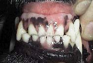
The close up shot of the gums of this normal dog are how healthy gums should look. Look at the detail of the gingiva (where the gums meet the tooth), and how adhered the gingiva is to the tooth.
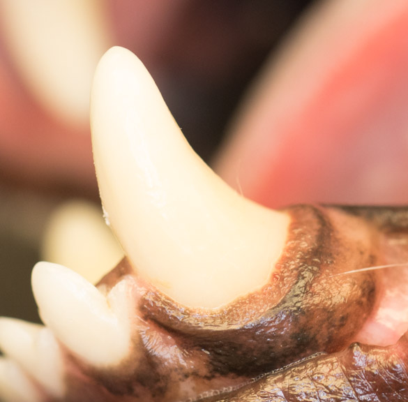
This is a healthy canine tooth with healthy gums
Stages of Periodontal Disease
There are 4 stages of periodontal disease:
Gingivitis only without loss of the periodontal attachment. Alveolar margins are normal. This gingivitis is reversible if the plaque causing this is removed.
Early periodontitis as evidenced by less than 25% attachment loss. This can be seen radiographically. This stage and the following stages are not easily reversed.
Moderate periodontitis where there is between 25% – 50% attachment loss. This is determined radiographically or by probing with gum line.
Advanced periodontitis with more than 50% attachment loss.
The first stage occurs when bacteria cause an invisible film of plaque to form on the teeth. The bacteria react with minerals and other debris that accumulate in the oral cavity, eventually causing tartar. You learned about his already in the biofilms section.

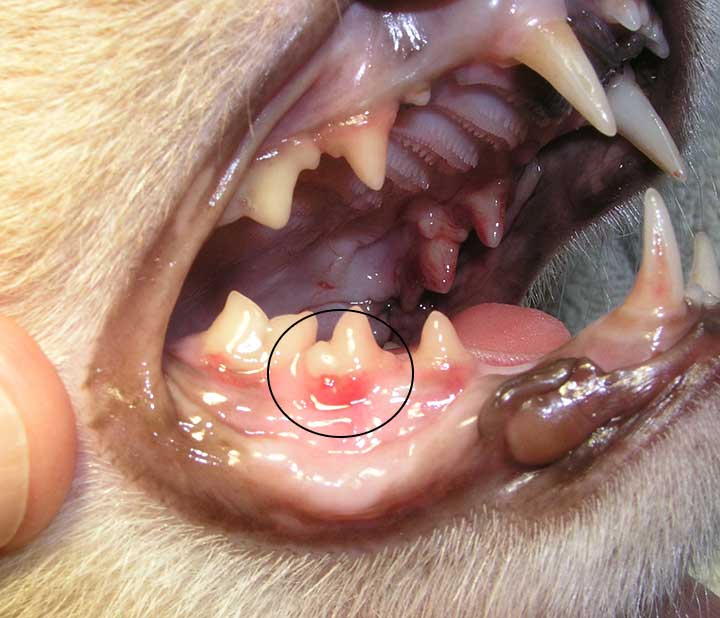
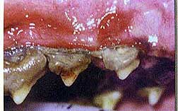
The lower canine tooth in this dog has tartar and gingivitis, as evidenced by the inflamed gum at the base of the tooth. This means bacteria are already causing trouble below the gum line

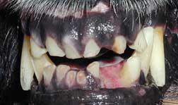
Look at the large upper tooth in the top middle of the picture with tartar. The red area of the pink gums just above this large tooth is gingivitis from bacteria. This gingivitis is significantly weakening the periodontal ligament and weakening the bone of the jaw.

This cat has the same problem, with gums that are even more inflamed
Left untreated, the teeth eventually progress to Stage IV periodontal disease. You saw a picture of a dog with problem at the beginning of this page. In Stage IV periodontal disease the tartar can be so extensive that it is the only thing holding the teeth in the socket in some cases. These dogs and cats cannot chew because the roots are rotten and it is painful. When we remove the tartar the teeth literally fall out.
The tartar is so thick that it is literally holding the teeth in place! It’s hard to believe that someone would let their dogs teeth progress this far. Unfortunately, this is not an uncommon situation.
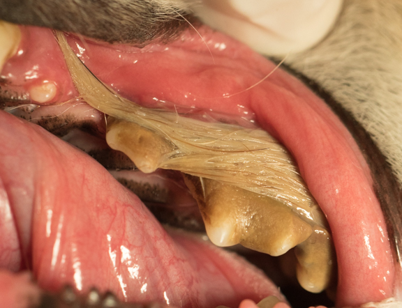
Some dogs that chew on their skin due to allergies will get the hair wrapped around their teeth and erode the gums

When we remove the hair the roots of the teeth are exposed due to the erosive nature of the hair on the gums.
The arrows point to the exposed roots. This is painful, and the teeth need to be removed.
In some cases the infection under the gum line has eroded away the gum tissue that normally covers the root. If the tooth has 2 roots it will cause a hole to appear between the roots where the gum has eroded- this is called a furcation lesion.
This is one potential outcome when pets with periodontal disease are not treated. The teeth in this cat literally rotted out of its mouth.
This situation is completely preventable. Fortunately, pets that have no teeth can still eat well, but that is small consolation for this cat. The years of chronic bacteria that were released into this cat’s bloodstream, when the periodontal disease progressed from Stage I to Stage IV, seriously affected the internal organs and caused this cat to have premature organ failure.

This is another potential outcome for a pet that has periodontal disease. This dog’s lower jaw (mandible) is fractured at the chin because of long-term periodontal disease. There are only two incisor teeth left (compare it to the picture below). A wire needs to be put in to hold this jaw together.
This is what the jaw and incisor teeth look like on a dog with healthy teeth

This is how we wire a jaw together to minimize pain and get this pet eating on its own again. It is wrapped all the way around the jaw and anchored under the chin. It will need to stay in place at least one month.
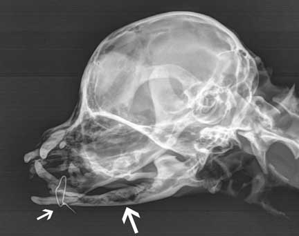
The left arrow points to the wire on this radiograph . The large right arrow points to the diseased mandible where we removed rotten teeth.
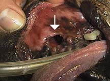
There are other serious complications that can occur when proper oral hygiene is neglected. This dog had a seriously infected tooth that created a fistula (arrow) into its upper jaw. Food will go into the passage and end up in the nasal cavity, which is not a place where food belongs. This dog will have chronic respiratory infections because of this, which can even lead to life-threatening pneumonia.
Heart Problems Secondary to Periodontal Disease
The heart is one of the internal organs that can be affected in advanced dental disease, because bacteria from the mouth infection can readily deposit on the heart valves (especially the mitral valve). Our heart page has extensive information if you would like to learn more.
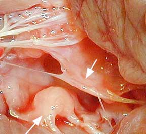
This picture is from our heart page. The top arrow points to a normal mitral valve leaflet. The bottom arrow points to a thickened mitral valve leaflet, which could be the result of chronic bacteria from the mouth. The thickened and rounded lower leaflet causes the problem.
The thickened valve can malfunction and leak blood backwards through one of the chambers of the heart, instead of forward like intended. This turbulence of blood as it flows through this leaky valve can often be heard as a heart murmur. The potential result of this back pressure is congestive heart failure- a buildup of fluid in the lungs (pulmonary edema). Fluid in the lungs will cause your pet to start coughing and feel very ill- it is a serious sign that requires immediate veterinary care.
In addition to heart (cardiac) problems, dental disease can affect the kidneys and the liver. These are both vital organs, and require a pet free from dental problems if they are to function properly. Some pets do not live a full life due to the chronic affect the bacteria has on their internal organs, leading to premature organ disease.
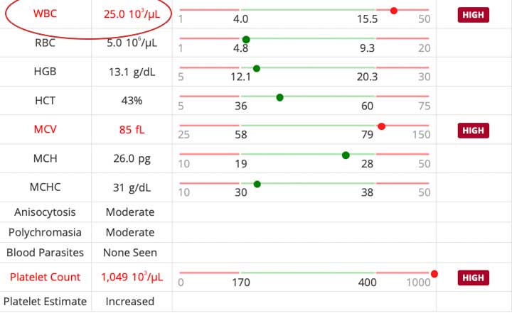
Chronic bacteria in the bloodstream from periodontal disease can cause the white blood cell count to elevate
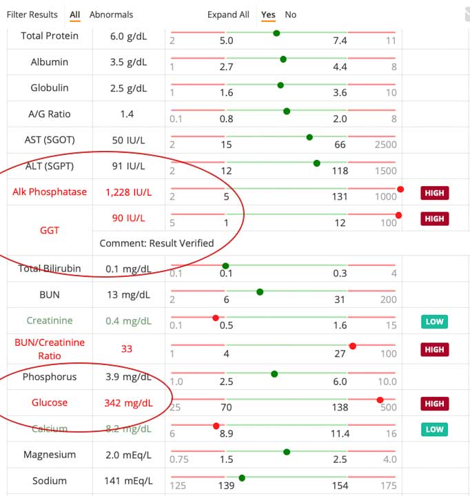
Chronic bacteria in the bloodstream from periodontal disease can cause the liver enzymes to elevate. If a pet has diabetes, like this one with an elevated blood glucose, the bacteria can make regulating the diabetes by using insulin almost impossible.
Diagnosis
As with any illness, the diagnostic process is carefully followed so that a correct diagnosis is made, and other problems that are a result of the dental disease (ex-heart murmur), or are occurring simultaneously (ex-kidney disease), are not overlooked. Since there are numerous diseases and conditions that can mimic dental disease, the diagnosis of dental disease must be performed by a veterinarian.
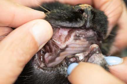
During a routine physical exam we will be be performing a complete examination, including the oral cavity. If dental disease is present, it is during this exam that we determine if your pet needs an anesthetic professional cleaning or a non-anesthetic professional cleaning. We can only perform a complete oral exam, looking at the tonsils, tongue, and back of the mouth, when your pet is anesthetized. This allows us to find oral foreign bodies, stomatitis, and cancers of the mouth, that we cannot see when your pet is awake.

We never would have diagnosed the severe stomatitis in the back of this cat’s mouth without an oral exam under anesthesia. This is a painful condition and not a diagnosis that should be delayed.
A complete oral exam is also performed during non-anesthetic dentals. Due to the thorough nature of the non-anesthetic professional cleaning, we sometimes find problems that require an anesthetic professional cleaning.
We will also show you how to perform a basic oral exam so that you can monitor your pet’s progress at home. The oral exam is not complete until we exam your pets mouth (the medical term for the mouth is oropharynx) under anesthesia. Only then can we check for tumors, ulcers, gum disease, foreign bodies, and infections, and enlarged tonsils.

A basic oral exam, which you can do at home, would have found this tumor on the gums long before it got this large
Pre-anesthetic Preparation
The first step in the process is yearly exams by one of our doctors, and more often if there is a medical problem of any kind or you pet is on chronic medication. Many people will have this yearly exam performed when their pet comes in for yearly vaccines. We will look inside the mouth and determine if any oral disease is present. If there is enough gum disease to warrant professional cleaning we will order a pre-anesthetic blood panel. We will also try to identify teeth that might need removal if there is an obvious problem.
This blood panel will let us know if your pet is ready for anesthesia, will check your pets health in general, and will allow us to assess any damage to the liver or kidneys from the chronic bacteria in the bloodstream. In addition, our doctors will sometimes recommend other tests prior to anesthesia. These tests commonly include radiographs of the lungs or abdomen, along with an electrocardiogram to assess the heart. Our pre anesthetic diagnostic tests page covers these tests in more detail.
Our doctor will analyze the results of the pre-anesthetic diagnostics tests and customize an anesthetic protocol for your pet. In many cases Intravenous fluids will be given prior to and during the professional cleaning. These fluids, when used in combination with pre-anesthetic tests, dramatically minimize the risk of anesthesia. As a final preparation prior to professional teeth cleaning one of our doctors might put your pet on antibiotics.
Anesthesia
When significant gingivitis is present proper dental care involves more than just scraping tartar off the teeth. Just scraping the tartar may temporarily make the teeth look better, but it is not addressing the real problem that occurs under the gum line. Thorough dental care involves scaling, probing, radiographing, flushing, measuring, fluoride and polishing. You will learn more about these in the next section.
These treatments can only be accomplished on an anesthetized pet. It is not realistic to think that all of this can be accomplished on an awake pet, and be as thorough as we need to be.
When these procedures are performed properly we can reverse the periodontal disease in some cases, and keep the teeth and gums healthier for a longer period of time. Since the risk of anesthesia is negligible with the precautions we take and the precise method available to administer and monitor anesthetic, it is well worth the negligible risk in order to clean the teeth and gums properly.
In reality, the risk of disease occurring by not cleaning your pet’s teeth professionally is greater than the risk of anesthesia.
We have extensive experience in anesthetizing pets, especially the geriatric pets that so commonly have advanced dental disease. To minimize any anxiety you have over anesthetizing your pet, one of our doctors will personally discuss our anesthetic protocol with you and set up an anesthetic plan that is specific for your pet’s condition. Our anesthesia page has extensive detail on how we anesthetize animals.
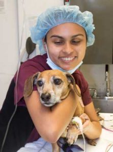
Our patients are giving plenty of TLC before we anesthetize them

We keep a close tab on important physiologic parameters for all of our surgeries. Monitors like this give us an early warning of an impending problem.
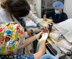
We have multiple people monitoring your pet when it is under anesthesia
Professional Cleaning
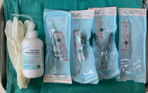
Before we clean any pet’s teeth we make sure all of our dental instruments are sterile and ready to go. Once we put a pet under anesthesia we do not waste any time getting organized, we get organized first.
This cleaning has four main components:
- Cleaning above the gum line with the ultrasonic scaler
- Cleaning under the gum line with special instruments
- Probing and examining of each tooth, with dental radiographs, to look for root decay and loss of bone
- Removal of rotten teeth
- Deep cleaning under the gum line with a curette, called root planing, to get at the bacteria and plaque in deep pockets
- Placing antibiotics on teeth with deep pockets in order to save them
- Antimicrobial medication to control the periodontal disease
- Oral sealants to prevent plaque buildup and the recurrence of the problem
Every pet is different, and we might do some or all of these procedures.
Oral Exam and Charting
The first aspect of the cleaning process is an examination of the complete oral cavity. It is only when a pet is sedated can this be completed thoroughly.
The arrow is pointing to a cyst in the mouth of this dog that was not seen until it was sedated. The owner did not know it was present, nor did this dog show any symptoms. We were able to remove it before it became a problem.
After our thorough oral exam we chart the problems encountered
Dental Unit
The equipment you will find in our hospital is the most advanced available. It allows us to provide a wide array of dental services.
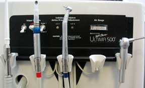
We use a specialized ultrasonic scaler that is made for animal teeth
Radiology
Radiography is an important part of dental care and is commonly performed as the next step after the oral exam. During your pet’s oral exam under anesthesia our staff will measure the depth of the pocket on the teeth that have disease. If the depth is 4 mm or greater we might take a radiograph of the tooth to make sure the underlying jawbone and root are healthy. If the root or jawbone are not healthy the tooth needs to be removed or a root canal at a specialist needs to be performed.

If there is a large pocket or bleeding occurs when we probe the tooth, we will radiograph it to see the roots
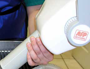
Our dental x-ray machine is made specifically to radiograph animals

The machine is automated, allowing us to rapidly take high quality radiographs
The dental X-rays are carefully analyzed by one of our doctors
The high definition of these radiographs allows us to see problems that are not apparent during the oral exam. Here is the normal tooth radiograph you saw at the beginning of this page.
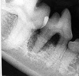
This radiograph shows a problem around the root. Do you see the dark, semicircular area around the root of the tooth in the very center of the picture? Compare it to the other root of this same tooth just to its right. This dark semicircular area radiographically is called lucency, and is an indication of deep seated infection in the tooth. It is painful and needs removing.

The red circle encompasses the problem root
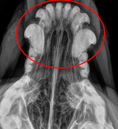
In this radiograph the jaw bone has been eroded down to expose parts of the roots on both teeth. This is the furcation lesion shown earlier.

Do you see the damage to the two teeth at the top right of this radiograph ?
Calculus Removal above and Below the Gum line
If the tartar is extensive, as it is with this dog, a special dental instrument is used to crack off large pieces of tartar before we use the scaler.
This enables us to clean the teeth faster, another method to minimize anesthetic time. It also reduces wear on the ultrasonic scaler tip.
Scaling teeth is greatly facilitated by a special instrument called an ultrasonic scaler, which you learned about earlier in this page. By vibrating tartar off the teeth with the scaler we cause minimal trauma to the tooth enamel. In addition, the rapid manner in which it removes the tartar minimizes anesthetic time. The gentle nature of the scaler allows us to clean under the gumline and not irritate the gums.

The tip vibrates 18,000 times per second, and literally vibrates tartar off the teeth. It does not harm the enamel, and lets us clean the teeth faster than doing it by hand. It continually sprays water to minimize heat buildup which could irritate the gums.

It has a special light to give us good visualization so we do not miss anything or harm the gums
Probing and Measuring

Here is a close-up of the probe
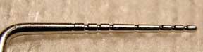
Each of the notches is 1 mm, the total length being 10 mm. Anything more than a 3 mm pocket under the gums in dogs, and 0.5 mm in cats, is significant.
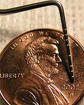
Lets have a little fun and show you just how small 10 mm is, courtesy of Uncle Abe

When we measured the depth of the pocket on this tooth it was obvious from the bleeding and the depth of the probe that periodontal disease is present and a radiograph is needed
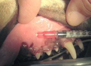
If we think the bone loss seen on the radiograph is manageable, and the gum pocket is not too large, we can place a long acting local antibiotic, called Clindoral®, under the gum line. This will continually kill the bacteria causing the gingivitis. The ultimate goal is to save the tooth from advanced periodontal disease and the need to remove a rotten tooth. If the problem is too advanced for this treatment, we will remove the tooth.

It contains an antibiotic called Clindamycin

It comes in a prepackaged syringe

It allows for precision placement of this antibiotic under the gums, with the long term goal of saving the tooth
In some pets the tooth problem is so severe that removal of the tooth is necessary. We make this determination only after probing, taking a radiograph, and exploring the option of using Doxirobe® or Clindoral®.
Before we extract a tooth we use a short acting and long acting local anesthetic, in addition to the general anesthetic currently being used. This nerve block allows us to use less general anesthetic.
After removal of a tooth we suture the gums over the opening for faster healing, and to prevent food particles from going in the socket.

This badly infected and painful canine tooth in this cat needs removal
This is the opening that remained after removing the tooth. The gums are hardened and the opening cannot be sutured closed.

If the opening cannot be sutured, we pack the opening with a special bone graft

This is the opening with the bone graft in place
Root Planing
For the remaining teeth, root planing is probably the most critical step in the professional cleaning process. By scraping the bacteria under the gum line with this special instrument we take care of the problem at its core. This can only be done on your pet when it is under anesthesia.
We use a specially designed instrument that is gentle yet thorough

Root planing allows us to get at those bacteria under the gum line
Flushing the Gums
After the teeth are scaled and probed, and the roots have been planed to remove the originating bacterial cause, we spray them with chlorhexidine to further help eliminate the bacteria that are causing gingivitis. It is only after this point in the professional cleaning process that we have significantly decreased those billions of bacteria.
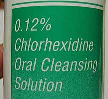
This special antibacterial reduces the bacteria burden in the mouth
Polishing
Polishing the teeth makes them look whiter. It also smoothes off the enamel surface and makes it more difficult for bacteria to adhere. Once bacteria get reestablished, the cycle of plaque leading to tartar and eventually gingivitis gets started all over again.

The teeth are polished with the same instruments dentists use on us
Fluoride Treatment
One of the final steps in the cleaning process is the application of fluoride to prevent cavities. We bathe the teeth in fluoride for a few minutes, then rinse it off. It has a very nice smell, too bad we can’t transmit smells over the Internet. We even put fluoride on the teeth of pets when they are spayed or neutered to help protect their teeth when they get older.

It smells good and is fun to watch as it foams up and covers the teeth
If the enamel on your pet needs a sealant, we use Oravet®. It significantly reduces plaque and tartar formation by creating an invisible barrier that helps prevent bacteria from attaching to your pet’s teeth.

It comes prepackaged with a special applicator

It is applied directly on the enamel
Prevention
Due to the short life span of pets in relation to people, proper home care of your pet’s teeth becomes an important health measure. Just like in people, routine preventive care is critical to proper dental hygiene. This was discussed at the very beginning of this page. This saves your pet from extended periods of pain and unnecessary tooth loss, and can save you the expense of the veterinary care needed to treat advanced dental disease.
Your pet’s teeth should be checked every 6-12 months by one of our doctors, especially if it has already had gingivitis and had its teeth cleaned. Any pet that has had periodontal disease should be checked every 6 months. One of these check ups can be accomplished when your pet is brought to our hospital for yearly booster vaccinations.
One of the most important things you can do to slow down the recurrence of dental disease is to brush your pets teeth. This will help keep the gums healthy and prevent tartar buildup on the teeth on the cheek side (buccal) of the mouth, although it does not work as well on the teeth on the tongue (lingual) side of the mouth. Even though this may sound like an impossible feat for an uncooperative pet, it is one of the best ways to prevent dental disease.
The teeth will eventually need professional cleaning again in the future (most people get their teeth cleaned several times per year). This is where non-anesthetic dentals come in to play. The teeth will be cleaned professionally, yet anesthesia is not needed. This is assuming you are doing some brushing or other dental care talked about at the beginning of this page. Proper brushing will decrease the amount of dental disease that occurs and the number of times we will have to clean your pet’s teeth over its lifetime.
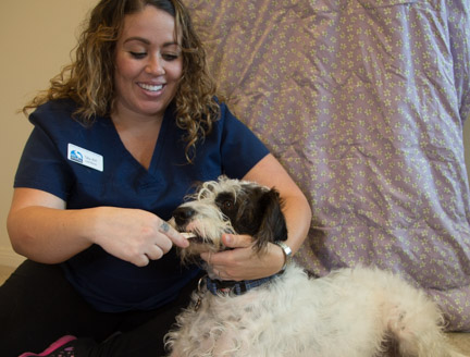
When brushing the teeth there are some common sense things to do to make the process go smoother. One of our technicians will demonstrate some of these techniques with one of our hospital cats (they love the attention). It is important to remain calm and patient, since for most pets having something put into their mouths is a new experience. With a little tincture of time, the procedure progresses smoothly. It is highly advantageous to start the brushing process at an early age.
Patience is the key! Try to do something positive (feeding it, playing or walking) with your pet just after brushing to condition the behavior for the future. Try to make the whole process fun, and don’t ever let on that you are doing something good for your pet (kinda like child psychology- if its good for them they won’t do it). With your pet near you or on your lap, maybe while watching TV, let your pet get used to your finger near its mouth.
Dipping your finger into a food or liquid your pet has acquired a taste for helps start the process smoothly. When it is comfortable with your finger, use a soft gauze to massage the gums and gently rub the teeth. a cotton tipped applicator can also be used. Eventually you want to progress to a toothbrush.
In smaller pets, especially cats, proper restraint is important. There needs to be a proper balance between too little and too much restraint, a balance that varies with each pet. This is especially true with cats. For smaller pets, placing them on a table will make the process go smoother. Larger pets can also be placed on a table, if feasible, or can be restrained on the ground. Only one or two people should be involved in the cleaning process, usually without children present. We have a complete page demonstrating this restraint technique.
Eventually, introduce a soft bristled toothbrush. These toothbrushes are available in our dental kits. A rubber finger brush can be used but a toothbrush is preferred. You should not use your personal toothpaste to brush your pet’s teeth because the taste can upset their stomachs. Our dental kit has toothpaste that is specially made to be palatable to animals. These kits also have suggestions to make it easier to brush your pets teeth.
If you consider daily tooth brushing a chance to enhance your bond with your pet, you and your pet will find it more enjoyable. Brush the teeth in a slow and circular motion with a small amount of toothpaste. It’s important to brush the outside of the teeth (the teeth up against the lips and not the teeth up against the tongue) since that is where the plaque is most prevalent. If your pet is cooperative brush the insides next. Your goal is to brush at least 3 times per week. This will decrease plaque by 90%.
If you encounter resistance on a pet that normally lets you brush, or see blood or there are blood tinges on the toothbrush, smell any odor, see any inflamed area or swelling, or a buildup of tartar or inflamed gums, you should bring your pet in for an exam. If the tartar is significant it is time for a professional cleaning.
In some cases brushing is just not feasible. In these situations you can use the dental treatments recommended at the beginning of this page.
Congenital Abnormalities
Small breed dogs tend to have dental problems more often than large breed dogs. This may be due to the fact that they have smaller oral cavities and the teeth are forced closer together. Cats get comparatively few congenital problems regarding their teeth. Any condition where the teeth are not normally positioned is called a malocclusion.
Malocclusions are corrected only if there is a problem with mastication (chewing). Undershot jaw (lower jaw protrudes beyond the upper jaw) is seen on occasion, and is prevalent in small dogs and in breeds like Bulldogs, Shih Tzu’s, and Lhasa apso’s. Overshot jaw (upper jaw protrudes beyond lower jaw) is similar to buck teeth in people.
Occasionally, a dog will not shed a deciduous tooth when a permanent tooth starts to come through the gums in the same location. These retained deciduous teeth, along with any extra teeth, should be removed because they will result in displacement of the permanent teeth. Problems of this nature are discovered by our doctors on routine exams. This enforces the importance of bringing in young pets for vaccines and exams at an early age.
Miscellaneous Dental Problems
Carnaissal Tooth abscess
The carnassial tooth (upper 4th premolar) may become infected and result in the formation of an abscess around the root. This is a very painful condition and is often accompanied by fever, loss of appetite and depression. a classic symptom of the problem is discharge through the face below the eye. This tooth needs to be removed for the problem to be corrected. It has a deep root and needs careful extraction to correct the problem.
Lymphoplasmocytic Gingivitis
This disease, seen almost exclusively in cats, is a specific inflammation of the the gum tissue. It is a painful and debilitating condition that is controlled but not cured. It is treated in various ways, including surgery with a laser.
The best way to treat this problem is to remove all of the teeth. Even though this seems like an extreme measure, the gums on these pets are so painful that removing is the only way to take away the pain. A cat with no teeth has no pain, and can eat well, compared to a cat that has all of its teeth and is in significant pain.
This picture from earlier in this page shows how inflamed the mouth can become with this problem. This needs to be biopsied to make sure it is not squamous cell carcinoma.
Cracked Teeth
It is very common for pets, especially dogs, to break or loosen their teeth while playing or chewing. This can cause significant discomfort and predispose your pet to dental problems later on in life. Injured teeth are usually removed, under general anesthesia, to ensure that the whole tooth is removed, including the root. If the root is not removed there will be a continual problem. In some cases we will refer you to a specialist that will determine if the tooth can be saved.
This dog fractured its tooth by chewing rocks
It was so badly traumatized it had to be removed to prevent pain and infection going into the root of this tooth. This tooth has 2 deep and strong roots, so it has to be split in half with a high speed drill.
The tooth just after removal
Worn Down Teeth
Worn down teeth are usually caused be chewing rocks, chains, and fences. This is a behavioral problem that should be corrected to prevent long term problems. also, dogs that continually chew or bite at the skin due to allergies or fleas will cause the incisor teeth to be worn down, sometimes all the way to the gum line.
This problem can be detected during a routine exam and corrected by prevention of chewing on itchy skin before the teeth get worn down too far
At this stage there is no way to correct the problem without extensive dental work with a specialist
Growths
Pets can get growths in the oral cavity, some of them can be benign, some are cancer and are malignant. Any growth or inflamed area in the mouth should be biopsied.
A benign gum growth that occurs usually in older dogs is called an epulis. The growth of the gum sometimes become so large that it covers a tooth almost completely.
This is the same growth as above 1 week after removal using the laser
What is the next step?
If one of our doctors feels your pet needs to have its teeth professionally cleaned there are several steps you should take:
Make an appointment to have the teeth cleaned before you leave our office. This will give you greater flexibility in your scheduling and allow us to accommodate you as much as possible. One of our receptionist’s will give you a written price estimate based on the doctor’s written instructions. An estimate will be given that covers all anticipated costs.
Even though our estimates are very accurate, there may be slightly greater, and more often lesser, charges on the final bill. This might be because some teeth need removal or medication needs to be sent home, or even finding something on the oral exam while under anesthesia that was not readily visualized during the initial exam. If there is any significant change in the price we will call you before proceeding. Please leave a phone number where you can be reached.
If one of our doctors feels your pet needs pre anesthetic diagnostic tests, have them obtained while you are here, or drop your pet off and return to pick it up later when the tests are complete. Any test samples sent out to our outside laboratory will be available the following morning. Please call our office after 10 AM the next day for these test results.
The night before the teeth cleaning take away all food before you go to bed. It is OK for your pet to drink water in the morning. Our office opens up at 7:30 AM for drop offs. We appreciate having your pet in for its teeth cleaning by 8 AM.
We will anesthetize your pet and clean its teeth sometime in the morning or afternoon. One of our doctors will call you as soon as the procedure is complete. It is very rare for a pet not to go home on the same day its teeth are cleaned. Your doctor will let you know if he plans on keeping your pet overnight. This might be because your pet is older or has a medical problem that requires us to monitor its progress in the hospital for an additional night.
The best time to pick up your pet is in the late afternoon or early evening. We are open until 10 PM every night for your convenience.You will be given written post dental instructions when you pick up your pet. If you have any questions after reading these instructions please let us know. Your pet may be groggy the first night. This is not because of the anesthesia, it is because of the pain injection many pets are given after their professional cleaning.
Contact with children and other pets should be supervised by an adult the first night. Give it a small amount of water and soft food an hour after getting home. If it eats and there is no vomiting, give it some more food and water. Please call us the next morning if you have any questions or you feel there is a problem.
If we send your pet home with pain medication or antibiotics use them exactly as prescribed.
Call us if your pet does not resume its normal activity and eating habits within 24 hours.